Collagen, the body’s most abundant protein, does not exist inside living cells as the rigid rod depicted in biology textbooks. Researchers in Barcelona have directly observed collagen behaving as liquid-like droplets, a discovery that may explain how cells safely transport this essential structural protein and could reshape research into fibrosis, wound healing, and cancer.
For more than half a century, cell biologists have wrestled with a fundamental question: how does collagen, one of the largest and most important proteins in the human body, travel through cells without causing damage?
Now, scientists at the Center for Genomic Regulation (CRG) in Barcelona believe they have found the answer. Their study, published in the Journal of Cell Biology, reveals that collagen exists inside living cells not as a long, rigid structure, but as a flexible, liquid-like condensate.
The discovery challenges a long-standing view of collagen biology and offers a new explanation for how cells handle the protein that forms the structural foundation of skin, bones, tendons, and organs.
Textbooks May Have Been Showing Only Part of the Story
Collagen accounts for roughly one-third of the total protein mass in the human body. Scientists have long understood that purified collagen appears under a microscope as a rigid rod that can reach lengths of up to 400 nanometers.
That understanding created a major problem.
Collagen is produced inside a cellular compartment called the endoplasmic reticulum (ER). Yet the vesicles traditionally believed to transport proteins through cells are typically only 60 to 90 nanometers in diameter.
The mismatch in size has puzzled researchers for decades.
According to senior author Vivek Malhotra, the new findings suggest the puzzle exists because scientists were looking at collagen in the wrong state. The familiar rod-like structure represents collagen after it has left the cell and assembled into fibers. Inside the cell, collagen behaves very differently.
Rather than remaining rigid, it forms liquid-like droplets that are highly flexible and dynamic.
Direct Evidence From Living Cells
The breakthrough came through high-resolution live-cell imaging of human hepatic stellate cells, liver cells known for producing collagen and driving tissue scarring during fibrosis.
Researchers focused on procollagen 1, the precursor that eventually matures into type 1 collagen, which makes up around 90% of the body’s total collagen.
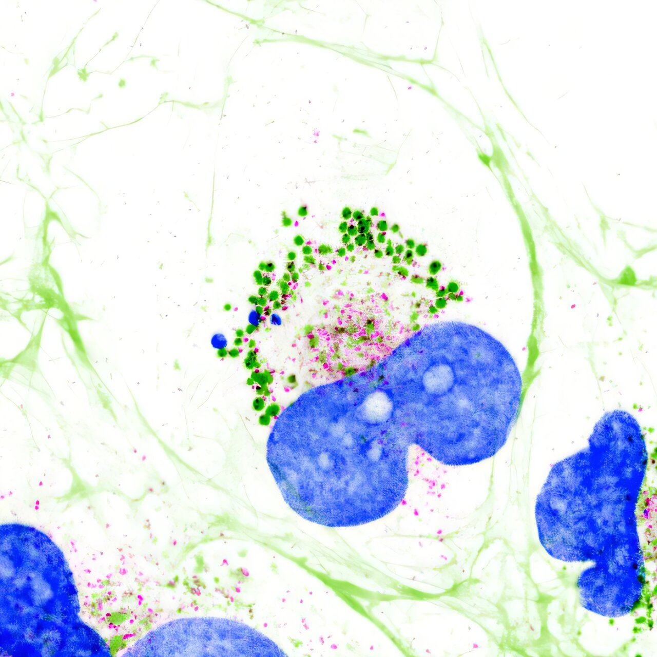
Inside these cells, the team observed collagen gathering into small spherical droplets. These structures merged together, split apart, and exchanged material with their surroundings.
Such behavior is characteristic of condensates, concentrated collections of proteins that separate from their environment in a manner similar to droplets of oil suspended in water.
The observations provide the first direct evidence of collagen’s natural state inside living cells.
Why a Liquid State Could Protect Cells
The discovery may also explain how cells avoid a potentially dangerous problem.
Collagen’s role outside the cell is to assemble into strong fibers that provide structural support to tissues. If that same fibrous assembly process occurred prematurely inside the cell, the consequences could be severe.
According to Malhotra, fibrous collagen forming within cells could be catastrophic and potentially lethal.
The liquid-like condensate state appears to offer a protective solution. By remaining fluid and pliable, collagen can be stored and moved through the cell without hardening into fibers before it reaches its proper destination.
In this view, the condensate acts as a built-in safeguard that keeps collagen functional while preventing cellular damage.
An Unexpected Discovery
The findings originated from experiments conducted by Soumya Bhattacharyya, a postdoctoral researcher in Malhotra’s laboratory.
In May 2024, Bhattacharyya was studying how collagen production increases in fibrotic liver cells when unusual spherical structures appeared in microscopy images.
The bright droplets immediately stood out.
At first, even the researchers were skeptical.
Because the observation challenged established cell biology concepts, the team initially suspected the structures might be artifacts or accumulations of defective protein.
To test that possibility, they examined whether the droplets were associated with BiP, a cellular chaperone involved in identifying and managing misfolded proteins.
The evidence pointed in a different direction.
Instead of showing signs of protein damage, the droplets contained helper proteins that recognize properly folded collagen. This suggested the structures were functional biological compartments rather than cellular waste.
Rethinking How Collagen Leaves the Cell
The study also sheds new light on TANGO1, a protein discovered by Malhotra’s laboratory about two decades ago and known to be essential for collagen export.
When researchers reduced TANGO1 levels, collagen droplets still formed. However, they no longer remained positioned at the ER exit sites where cellular cargo normally leaves the compartment.
As a result, collagen secretion declined.
This finding suggests TANGO1 may not function as a conventional cargo receptor. Instead, it appears to act more like an anchor, holding collagen condensates at the correct location for export.
Based on these observations, the researchers propose a new “liquid extrusion” model.
Rather than being packaged into traditional transport vesicles, collagen condensates may move through cellular exit sites using physical processes similar to wetting or capillary flow.
The researchers are still testing this idea, and direct visualization of the export process remains an important next step.
Potential Implications for Fibrosis and Cancer
If future experiments confirm the liquid extrusion model, the implications could extend far beyond basic cell biology.
Excess collagen production is a defining feature of many diseases, including fibrosis affecting the liver, lungs, and skin.
Collagen also plays a major role in cancer. Tumors often surround themselves with dense extracellular matrices rich in collagen and related proteins. These protective structures can make tumors harder for therapies and immune cells to penetrate.
The new findings suggest that proteins such as TANGO1 or the collagen condensates themselves could become potential targets for future research aimed at controlling excessive collagen secretion.
Researchers propose that disrupting TANGO1 or dissolving collagen condensates could be strategies worth investigating as scientists seek new ways to limit harmful collagen accumulation.
Why This Matters
This study overturns a long-standing assumption about one of the most abundant proteins in the human body. By showing that collagen exists as a liquid-like condensate inside living cells, researchers may have finally solved a biological mystery that has persisted for 60 years.
Beyond answering a fundamental question about cell function, the discovery opens new avenues for understanding how collagen is transported, how fibrotic diseases develop, and how tumors build protective barriers. While key questions remain, the findings provide a fresh framework for exploring some of the most important processes in human health and disease.