Beneath the sterile, cool glow of a modern medical facility, a series of ancient travelers from the Nile Valley are finally revealing secrets they have held for over two millennia. For decades, these Egyptian remains sat in museum collections, their internal stories obscured by the very bandages intended to preserve them for eternity. Traditional medical scans could only see so far through the dense, multilayered wrappings and desiccated tissue. Now, a breakthrough in imaging technology at Semmelweis University is peeling back those layers digitally, offering a high-definition window into the lives, deaths, and preservation techniques of the ancient world.
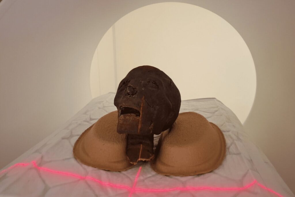
At the university’s Medical Imaging Center (OKK), researchers have begun a sophisticated diagnostic journey using a state-of-the-art CT scanner equipped with a photon-counting detector. This isn’t a typical hospital check-up; the subjects are archaeological finds from the Hungarian National Museum Public Collection Center (MNMKK) Semmelweis Museum of Medical History. By applying cutting-edge clinical tools to these fragile specimens, scientists are capturing highly detailed images that were previously impossible to obtain, promising a significant leap forward in our understanding of ancient Egyptian pathology and funerary customs.
Night Shifts and High-Tech Detectors
The logistics of scanning thousand-year-old remains require a delicate balance between modern healthcare needs and historical preservation. To ensure no disruption to living patients, the examinations are conducted deep into the night, outside of standard clinical hours. This allows the research team, led by Dr. Ibolyka Dudás, Chief Clinical Physician at the Department of Radiology, to utilize the full power of the institution’s newest imaging hardware.
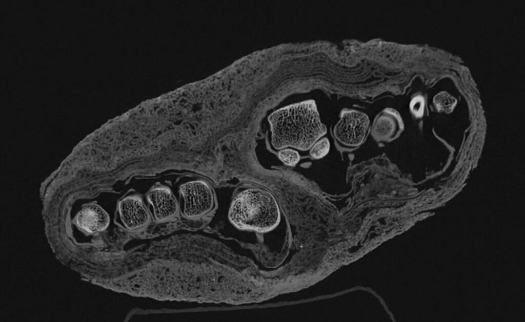
The photon-counting detector technology is the centerpiece of this study. Unlike conventional CT scanners, this system is exceptionally effective at analyzing complex, multilayered materials. For mummified remains, which often consist of skin, bone, resin, and multiple tiers of textile bandages, this precision is vital. The goal is to create the most accurate map possible of the internal structures, identifying any biological abnormalities and the specific chemical or physical preservation techniques used by ancient embalmers—all without disturbing a single thread of the original find.
Solving Centuries-Old Mysteries of Identity
The collection at the Semmelweis Museum of Medical History has been part of the institution since its founding, yet much of its history remained speculative. Previous attempts to study these finds included multidisciplinary examinations and conventional CT scans, but the technological constraints of the time left many questions unanswered. Even radiocarbon (C14) dating struggled with the samples; out of six specimens tested, only three yielded measurable results.
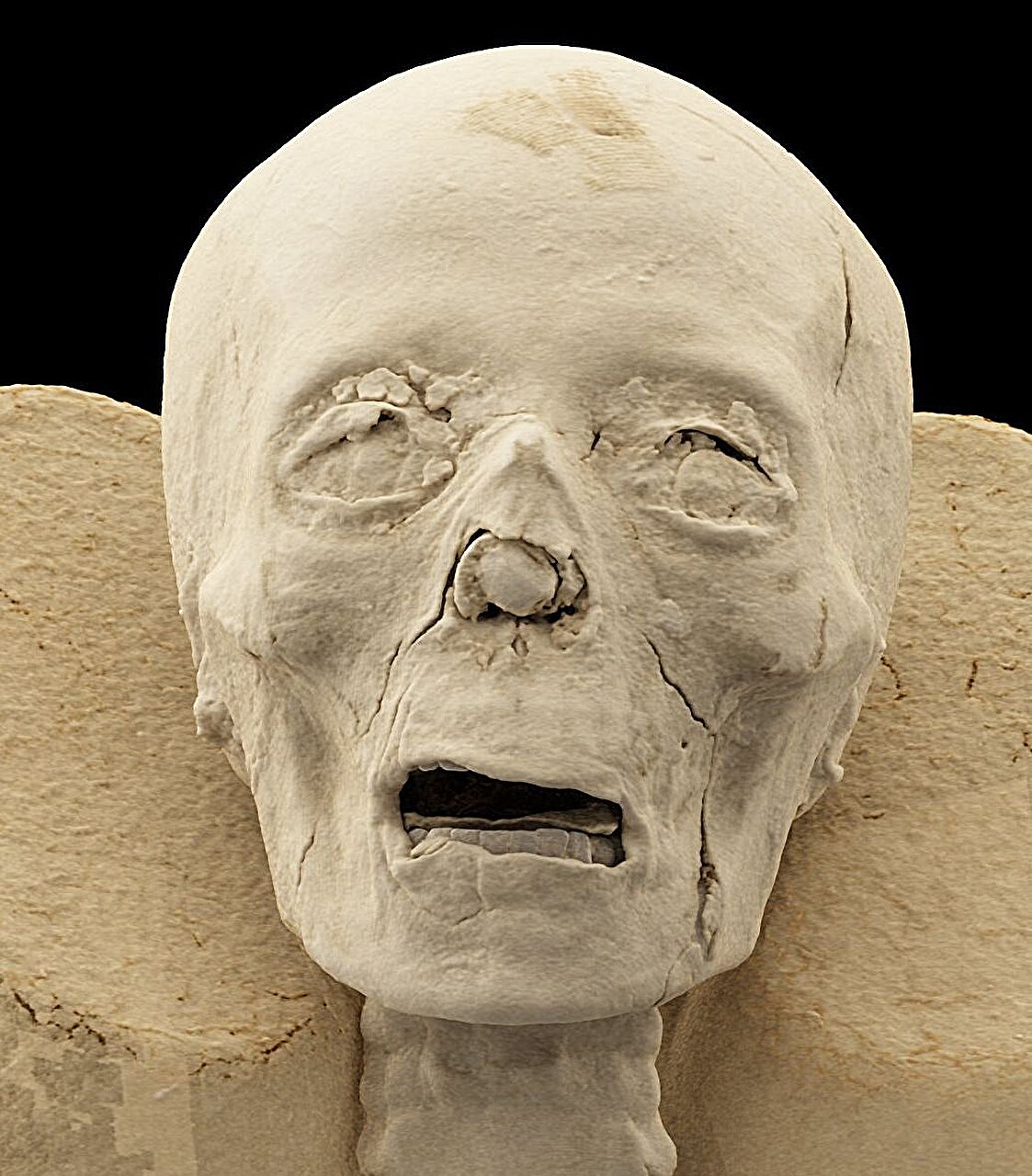
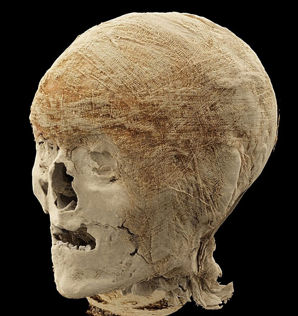
These successful tests, however, provided a crucial temporal anchor. The oldest remains in the collection date back to a window between 401 and 259 BCE, placing them at over 2,300 years old. With this chronological context established, the current series of high-resolution scans is focusing on two mummified heads to refine our understanding of who these people were. By examining the teeth and skull sutures in unprecedented detail, researchers can determine the age of the individuals at the time of death with far greater accuracy. These scans also serve as the digital foundation for future 3D reconstructions, potentially allowing for high-precision facial recreations that could show us the faces of these ancient individuals for the first time in millennia.
From Bird Mummies to Human Feet
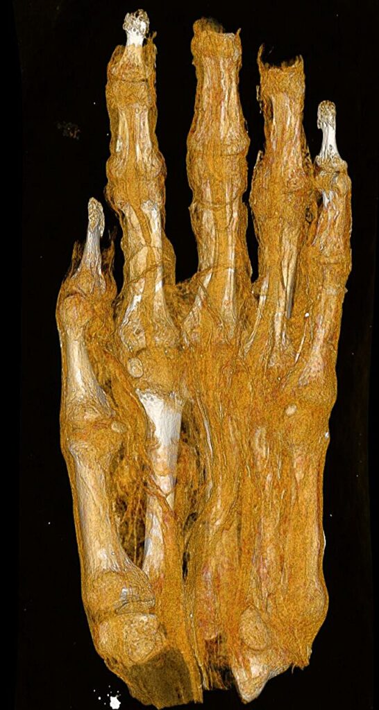
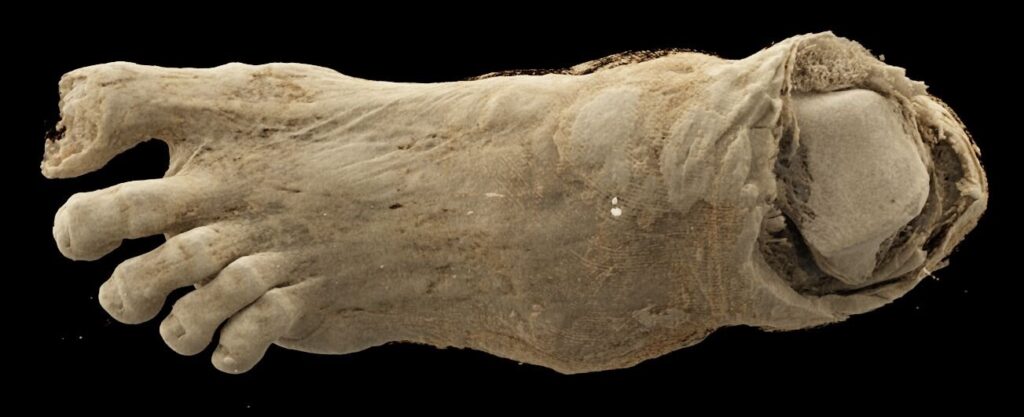
One of the most remarkable aspects of the new imaging project is its ability to correct long-standing historical misidentifications. In the world of archaeology, things are not always what they seem from the outside. One particular set of remains arrived at the museum identified only as a “mummy bundle.” Without the benefit of internal imaging, it was initially cataloged as a human head. Later, some hypothesized it might be the mummy of a bird.
It was only through CT technology that the truth emerged: the bundle actually contains a mummified adult foot. The new high-resolution scans are now going a step further, analyzing the distinct layers of the bandages wrapped around the limb. The images clearly show different structural characteristics within the textile remnants, which may provide clues about the specific mummification techniques used and whether the individual suffered from any identifiable illnesses. While researchers believe the foot was originally part of a complete mummy, the reason it was separated and the timing of that dissection remain mysteries that the team is still working to solve.
Diagnosing Ancient Ailments
The medical utility of the photon-counting CT is perhaps most evident in the study of a left lower limb that had previously stumped investigators. Older scans were unable to provide a definitive diagnosis for visible irregularities in the bone. However, the new, clearer images have opened up several possible interpretations. Current data suggests the individual may have suffered from osteoporosis, a condition characterized by weakened bones.
The challenge now lies in determining the “why” behind the diagnosis. Scientists are currently analyzing the data to decide if the bone loss was a result of natural age-related factors or a specific pathological process. Similarly, the examination of a second lower left limb has revealed that it likely belonged to a young individual. This marks the first time such detailed imaging data has been available for this specific find, allowing for a much more nuanced investigation into the life of a person who lived over two dozen centuries ago.
Why This Matters
This research represents a pivotal shift in how we interact with the past. By using non-destructive, high-precision medical technology, we can extract an immense amount of biological and historical data without compromising the physical integrity of the artifacts. For the curator of the collection, Chief Museologist Krisztina Scheffer, the project is the culmination of decades of preservation work. The ability to see “hidden” information within finds that have been in museum cabinets for years validates the importance of revisiting old collections with new eyes.
Ultimately, these scans do more than just identify diseases or age; they humanize history. By identifying the sex, age, and health struggles of these individuals—whether it is an adult with osteoporosis or a child whose hand is being measured for developmental markers—we move beyond viewing mummies as museum objects and begin to see them as people. This technological bridge between modern medicine and ancient history provides a more detailed view of human life and death than ever before, ensuring that the stories of these 2,300-year-old individuals are finally told with scientific validity and clarity.