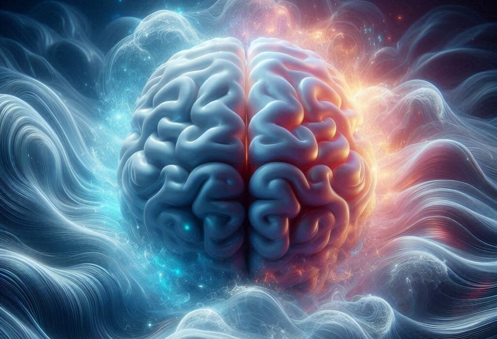
Deep within the folds of the brain lies a small, seahorse-shaped structure called the hippocampus. Though modest in size, it plays a central role in who we are. The hippocampus helps us form and recall memories, guides our learning, supports decision-making, and regulates emotional balance. Without it, the story of our lives would unravel—we would lose not only the ability to remember yesterday but also the capacity to imagine tomorrow.
For decades, scientists have studied this region to understand how its cells work together to shape cognition and mood. Yet the hippocampus has always been something of a mystery. We knew it contained diverse cell types—each with unique shapes, functions, and connections—but their precise identities and how they were arranged in the brain remained largely unmapped.
Today, thanks to groundbreaking advances in molecular biology and computational science, researchers have taken a leap forward in revealing the hippocampus’s inner workings.
The Tools of Modern Brain Cartography
Studying the brain is a challenge unlike any other. A single human brain contains around 86 billion neurons, and within the hippocampus alone exist countless subpopulations of specialized cells. To map this diversity, scientists need methods that can capture the fine detail of gene expression while also preserving the “geography” of the tissue.
Two powerful technologies have made this possible.
The first, spatially resolved transcriptomics (SRT), measures which genes are active in cells while preserving their original positions within brain tissue. Imagine not only cataloging the words in a book, but also knowing exactly where each word sits on the page—that’s what SRT does for the brain.
The second, single-nucleus RNA sequencing (snRNA-seq), zooms inside individual cell nuclei to study their RNA molecules. This allows scientists to classify cells into distinct subtypes based on their genetic signatures, even if those cells look similar under the microscope.
By combining these two approaches, researchers can not only identify the molecular identity of each cell but also see how these cells are arranged, layer by layer, within the hippocampus.
Building a Molecular Atlas
A team from Johns Hopkins Bloomberg School of Public Health, the Lieber Institute for Brain Development, and Johns Hopkins School of Medicine recently harnessed both SRT and snRNA-seq to produce the most detailed map of the human hippocampus to date. Their findings, published in Nature Neuroscience, represent a milestone in neuroscience.
The researchers studied hippocampal tissue taken from 10 adult donors who had no diagnosed brain conditions. They first collected vast amounts of molecular data, then used advanced computational methods—including an algorithm called non-negative matrix factorization (NMF)—to integrate the two datasets. This process allowed them to track gene expression patterns across cell types while preserving their precise locations in brain tissue.
What emerged was a comprehensive molecular atlas of the hippocampus, a high-resolution guide to its diverse cell subtypes and their spatial organization.
Revealing Hidden Patterns of Brain Activity
This atlas did more than just list cell types. It revealed how certain cells specialize in excitatory or inhibitory signaling—the balance of which underpins learning, memory, and emotional regulation. It also showed that the hippocampus is not uniform; instead, distinct regions such as the subiculum and presubiculum have their own molecular “signatures.”
To deepen their insights, the researchers compared their human data with equivalent maps from the mouse hippocampus. This cross-species analysis highlighted both similarities and subtle differences, shedding light on how evolutionary changes may have shaped human cognition. The comparison also suggested patterns of activity-dependent transcription, hinting at how hippocampal circuits rewire themselves during learning and memory.
Why This Matters
At first glance, this may sound like a technical achievement only relevant to specialists. But its implications ripple far beyond the laboratory.
The hippocampus is at the heart of many brain disorders, from Alzheimer’s disease and depression to epilepsy and schizophrenia. By creating a detailed reference atlas of its cell types and spatial organization, scientists now have a powerful tool for future research.
This resource could help researchers identify which cells are most vulnerable in disease, why some circuits break down, and how therapies might be targeted more precisely. It also opens the door to new questions about how the healthy brain supports memory and emotion—and what happens when those processes falter.
An Interactive Map for the World
Importantly, the atlas created by Jaqueline R. Thompson, Erik D. Nelson, and their colleagues is not locked away in a lab. The team has made both the dataset and an interactive online platform available to the global scientific community. This means researchers everywhere can explore the molecular geography of the hippocampus, accelerating discoveries that may one day transform medicine.
A Deeper Understanding of Ourselves
The hippocampus is more than a structure inside the brain—it is the seat of our lived experience. It is where memories are stitched together, where knowledge is consolidated, and where emotions find balance. To map it is to glimpse the architecture of our humanity.
With this new molecular atlas, we stand at the threshold of deeper understanding. Each cell type, each connection, each gene expression pattern is a clue to how the brain creates thought, memory, and emotion. Science has not yet unlocked all its secrets, but step by step, we are illuminating the labyrinth.
And perhaps, as we chart the hippocampus more clearly, we will also chart ourselves—our past, our hopes, our resilience, and the fragile, wondrous machinery that makes us human.
More information: Jacqueline R. Thompson et al, An integrated single-nucleus and spatial transcriptomics atlas reveals the molecular landscape of the human hippocampus, Nature Neuroscience (2025). DOI: 10.1038/s41593-025-02022-0.