For centuries, the question of what gives rise to conscious experience has fascinated philosophers, scientists, and laypeople alike. Is awareness born solely in the ripples of our cerebral cortex, or does it reach deeper into the brain’s inner sanctums? A groundbreaking new study led by researchers at Beijing Normal University provides compelling evidence for the latter.
The team has uncovered a hidden network of high-order thalamic nuclei—buried deep in the brain—that appear to act as gatekeepers to human conscious perception. Their findings, published in Science, could rewrite prevailing models of awareness and may open fresh avenues in neuroscience, medicine, and even artificial intelligence.
Beyond Wakefulness: Decoding Conscious Experience
Consciousness isn’t a single phenomenon but a layered construct. On one hand, it’s the general state of being awake, alert, and responsive—what scientists call arousal. On the other, it’s the precise moment-to-moment tapestry of thoughts, feelings, and sensory impressions that make up subjective experience.
While much of the research into consciousness has zoomed in on the cerebral cortex—the brain’s outermost layer responsible for reasoning, memory, and voluntary movement—there’s a growing sense that this might be only half the story. Deeper brain regions like the thalamus have often been dismissed as mere relay stations for incoming sensory information. But what if these central structures were more than just middlemen?
The new study suggests that certain parts of the thalamus don’t just pass messages—they might be speaking the first words of awareness.
The Subcortical Blind Spot
Despite their strategic placement and intricate connectivity, subcortical structures such as the thalamus have remained underexplored in the consciousness conversation. This is partly because functional imaging technologies like fMRI and PET scans, while valuable, lack the speed and resolution to capture the rapid, fleeting dance of neural signals occurring within these deeper brain regions.
This technological blind spot has made it difficult to track how fast-changing sensory stimuli transform into conscious impressions. Are these subsecond transitions driven by cortical interpretation, or do they originate from elsewhere?
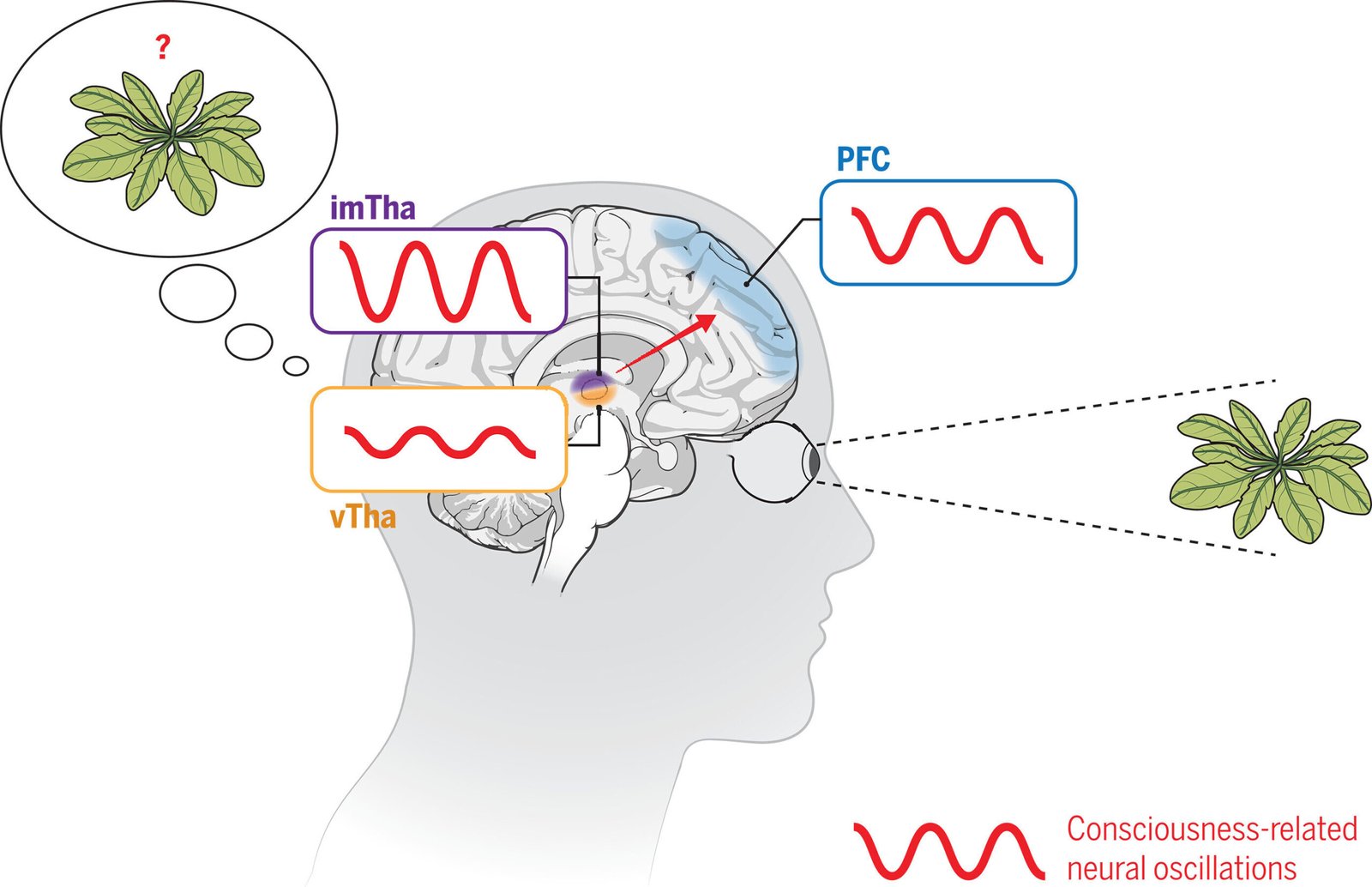
To answer this, the Beijing Normal University-led team turned to a powerful but rarely used method: stereoelectroencephalography (sEEG)—a technique that involves implanting electrodes directly into the human brain.
A Rare Glimpse Inside the Living Brain
The study took advantage of a unique clinical opportunity. Five patients suffering from chronic, drug-resistant headaches were undergoing invasive monitoring to prepare for deep brain stimulation therapy. With their consent, researchers placed electrodes in multiple thalamic nuclei and areas of the prefrontal cortex—allowing simultaneous recordings across 407 distinct brain sites.
The patients participated in a visual awareness experiment. They were shown barely visible black-and-white grating patterns, flashed briefly in peripheral vision. Participants indicated whether they perceived the stimulus by making subtle eye movements. By focusing on visual cues—rather than sound or touch—the researchers minimized interference from motor commands or language processing, isolating the pure experience of visual awareness.
The Spark Before the Flame
The data revealed something remarkable: activity in the intralaminar and medial thalamic nuclei consistently preceded the cortical signals associated with conscious perception. These deep brain regions lit up dozens of milliseconds before their cortical counterparts.
This wasn’t a random fluctuation. In 108 thalamic recording sites, the team found statistically significant differences in activity between trials where patients were aware of the stimulus and those where they were not—even when the physical properties of the stimulus remained identical. In other words, the difference was not in the eyes, but in the brain’s interpretation.
Among the earliest responders were the central medial, mediodorsal medial, and parafascicular nuclei—regions previously overlooked in studies focused solely on the cortex. These nuclei exhibited not only higher amplitudes in their electrical signals but also stronger bursts of low-frequency brainwaves during moments of conscious awareness.
A Symphony of Rhythms
To understand how these thalamic signals orchestrated consciousness, the researchers examined the rhythmic coordination of brain activity—particularly in the theta band (2 to 8 Hz), a frequency range associated with memory and attention.
Using phase synchrony analysis, they found that these theta waves originated in the thalamus, then traveled outward through the thalamofrontal loop, ultimately reaching the prefrontal cortex. This cascading wave of electrical activity formed a temporal choreography, hinting at a coordinated process rather than a scattered reaction.
In conscious trials, the low-frequency rhythms in the thalamus also modulated higher-frequency signals in the cortex through a process known as phase-amplitude coupling. This cross-frequency interaction didn’t just occur—it originated in the thalamus and was stronger and earlier than similar cortical activity. It’s as if the thalamus was conducting a neural orchestra, setting the tempo before the cortex began to play.
The Thalamofrontal Loop: A Conscious Highway
When researchers compared brain activity across different trial types, it became clear that thalamofrontal loop activity most accurately predicted conscious awareness. It outperformed all other variables—stimulus brightness, behavioral cues, or even eye movement direction.
Even in borderline cases where the stimulus was just below the perceptual threshold, the thalamic activity correctly distinguished conscious from unconscious trials. These findings suggest that the high-order thalamic nuclei don’t merely relay information—they initiate and coordinate the brain-wide activity that gives rise to perception.
Rewriting the Brain’s Map of Consciousness
This study delivers some of the most direct human evidence yet in support of a bold idea: that consciousness does not begin in the cortex. Rather, it may arise from thalamic seeds that bloom into cortical awareness through tightly timed electrical loops.
These findings resonate with emerging theories such as the Thalamocortical Loop Hypothesis and Integrated Information Theory, both of which argue that consciousness is not localized but distributed, emerging from dynamic patterns of connectivity and feedback between brain regions.
Importantly, the results challenge dominant frameworks that place the cerebral cortex at the epicenter of awareness. While the cortex undoubtedly plays a major role in interpreting and responding to stimuli, it may not be the birthplace of perception itself.
Implications for Medicine and Technology
By identifying the thalamic origins of consciousness, this research could have far-reaching implications for how we approach disorders of consciousness, such as coma, vegetative states, or locked-in syndrome. Targeted stimulation of high-order thalamic nuclei might one day restore awareness in patients trapped in unresponsive bodies.
The findings also offer fresh inspiration for the development of brain-machine interfaces. If conscious perception can be tracked or even initiated via the thalamus, future neuroprosthetics may tap directly into these loops to enhance sensory feedback or cognitive control.
Moreover, understanding how perception flickers into awareness could inform efforts to create artificial consciousness in machines—not just mimicking the outputs of cognition, but understanding the inputs that allow it to emerge.
Looking Forward: Lighting the Mind from Within
The discovery that consciousness may be sparked by a deep-set group of brain nuclei—quietly igniting before the cortex catches on—adds a profound new layer to our understanding of the mind. It reminds us that the brain is not a collection of isolated parts, but a living network of rhythms, signals, and circuits dancing in coordination.
In peering into the hidden rhythms of the thalamus, scientists may be getting closer to answering one of humanity’s oldest questions: How does the brain create the feeling of being alive? With every new insight, the mystery of consciousness becomes not simpler, but richer—and infinitely more fascinating.
Reference: Zepeng Fang et al, Human high-order thalamic nuclei gate conscious perception through the thalamofrontal loop, Science (2025). DOI: 10.1126/science.adr3675