For centuries, scientists have dreamed of seeing inside the human body without cutting it open—peering into the delicate networks of the brain, the pumping chambers of the heart, or the fine structures of other organs in their natural state. Imagine gazing through tissues as if they were glass, watching life unfold at the microscopic level without disturbing it.
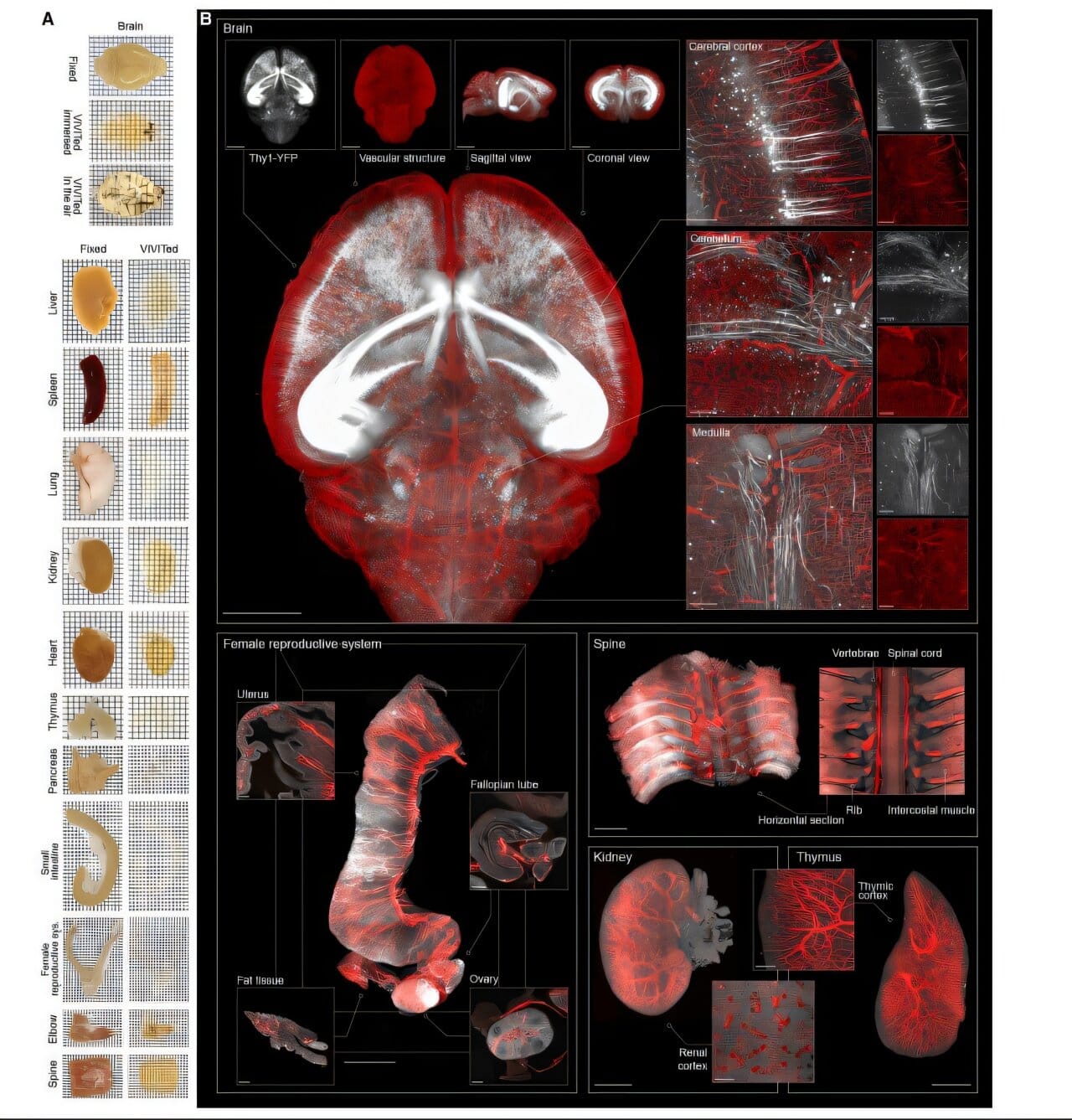
That dream has taken a leap toward reality. A team of Chinese researchers has introduced a groundbreaking technique that transforms biological tissue into a transparent, glass-like state. Published in the journal Cell, their study describes how this new method, called VIVIT, allows scientists to visualize entire organs at an unprecedented level of detail—without a single incision.
What Is VIVIT?
The method’s full name is a bit of a mouthful: vitreous ionic-liquid-solvent-based volumetric inspection of trans-scale biostructure, or VIVIT for short. At its heart lies an unusual set of materials called ionic liquids (ILs)—salts that remain liquid even at relatively low temperatures.
When tissues are treated with these ionic liquids, something remarkable happens. The samples undergo vitrification, a process where they become glass-like rather than forming destructive ice crystals. The result is a perfectly preserved, transparent organ that can be examined with high-resolution imaging tools.
This means researchers can now look at tissues in their true three-dimensional state—from whole organs down to the tiniest cellular connections—without cutting them apart.
Why Transparency Matters
To truly understand complex biological systems, scientists need to see not just cells in isolation but how they interact within entire networks. Traditional methods of studying organs often involve slicing tissues into thin sections and then piecing together a 3D reconstruction. But this comes at a cost: slicing can distort or destroy delicate structures, making it nearly impossible to rebuild an accurate picture.
Existing “optical clearing” techniques have tried to solve this by making tissues transparent, but most rely on organic solvents or aqueous solutions. These substances can cause tissues to shrink or swell, warping their natural architecture. VIVIT, however, avoids this pitfall by stabilizing the tissue in a vitrified, glass-like state.
This non-destructive approach preserves the organ’s original form and function while still allowing scientists to illuminate its hidden inner workings.
Mapping the Brain’s Hidden Highways
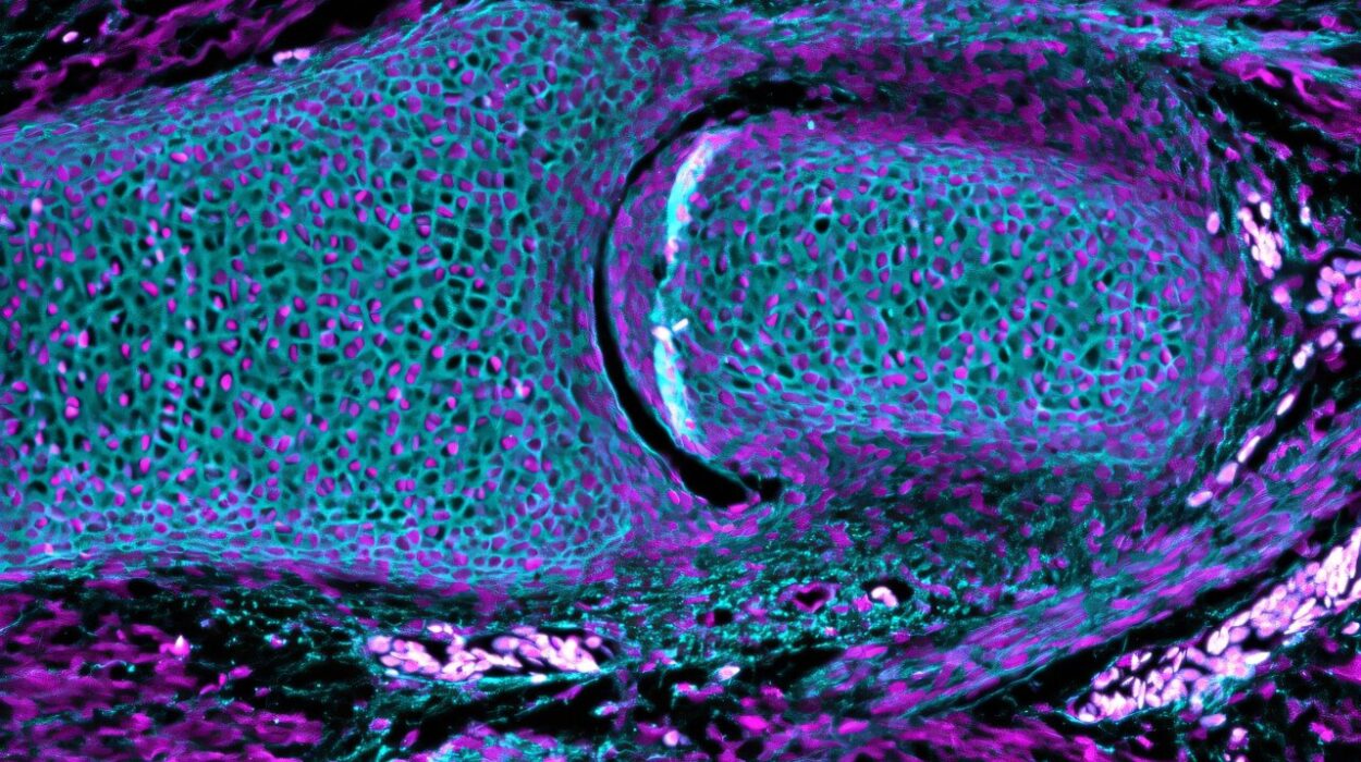
One of the most exciting demonstrations of VIVIT was in the brain. Using this technique, researchers mapped how multisensory neurons in the thalamus connect to their incoming signals and project their outputs across the brain. They also uncovered new insights into how inhibitory control functions in the cortex—a key process for balancing the brain’s constant electrical chatter.
By making the brain optically clear, VIVIT opened a window into the intricate wiring that underlies sensation, movement, and thought. Instead of seeing fragments, scientists could now observe the full continuity of neural highways.
Brighter Dyes, Clearer Views
As if making tissue transparent weren’t enough, VIVIT revealed another unexpected bonus: it boosted fluorescent dye signals. In some cases, brightness increased up to 38-fold. This is a game-changer for imaging, since fluorescent dyes are the main tools scientists use to label and track specific cells and molecules. With VIVIT, even faint signals become clear, enhancing the resolution of microscopy without additional intervention.
A Marriage of Biology and Software
To complement the physical clearing process, the researchers also created a digital partner: TARRS (Trans-scale Acquisition, Reconnection, and Reconstruction System). This software stitches together slices of tissue into seamless 3D models, preserving structural accuracy. Together, VIVIT and TARRS provide both the biological transparency and the digital reconstruction needed to study organs as never before.
Preserving While Exploring
Perhaps the most remarkable quality of VIVIT is its ability to both reveal and protect. Because the tissues are vitrified rather than chemically damaged, researchers can still return to the samples for additional tests or precise sectioning. This flexibility means scientists don’t have to choose between preservation and exploration—they can have both.
The implications extend far beyond neuroscience. From mapping heart fibers that coordinate each beat to tracing cancer cell invasion across tissues, VIVIT could become an essential tool for unraveling the mysteries of life at every scale.
Challenges and Future Directions
As revolutionary as VIVIT is, many questions remain. How exactly do ionic liquids interact with tissues to induce this glass-like transformation? Why do they amplify fluorescence so effectively? Unlocking these secrets could pave the way for even more powerful versions of the technique, possibly tailored for different tissues or imaging methods.
What’s clear, however, is that VIVIT represents a turning point. By making organs transparent, it offers not just a technological advance but a philosophical one: a reminder that much of biology’s beauty lies hidden in the layers we cannot ordinarily see.
A Window Into Life Itself
Science has always been a quest to look deeper, to peel back the layers of mystery surrounding life. From the invention of the microscope to the decoding of DNA, each breakthrough has revealed worlds once thought invisible. VIVIT now joins this lineage, offering a glass-like window into the human body.
For the first time, we can hold an organ in hand, look through its transparent layers, and witness the astonishing architecture that sustains life—without harm, without loss, without shadows. It is as if biology has finally given us permission to see itself clearly.
And in those clear tissues, glowing under the microscope, lies not just science but wonder—the breathtaking beauty of life revealed as never before.
More information: Yixiao Gao et al, VIVIT: Resolving trans-scale volumetric biological architectures via ionic glassy tissue, Cell (2025). DOI: 10.1016/j.cell.2025.07.023