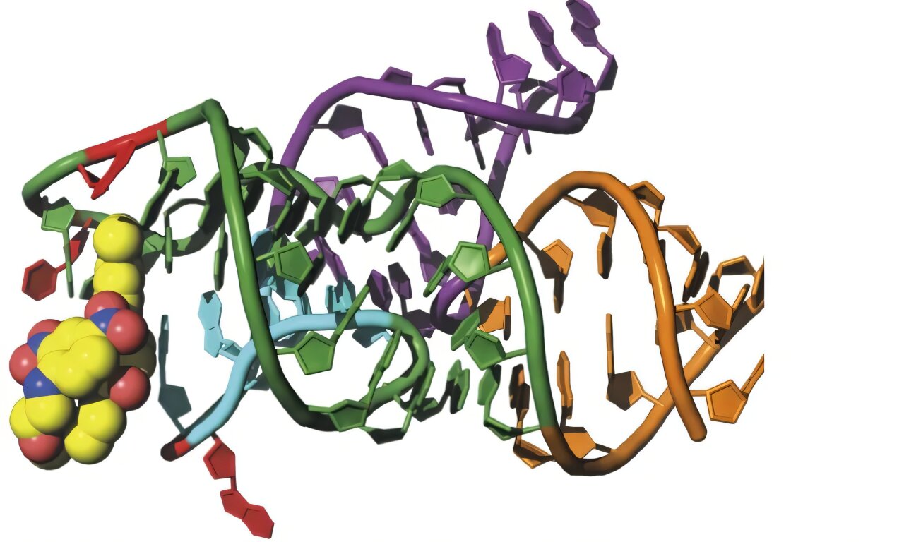
Inside a compact RNA structure, one nucleotide changes position as a fluorescent dye approaches, opening room for the dye to enter and helping turn on the fluorescence needed to track RNA at super-resolution. The nucleotide, G38, sits in a tightly packed region of the RhoBAST RNA aptamer when no dye is bound. When the fluorophore TMR-DN binds, G38 flips outward and stacks above another nucleotide, A37. That small structural movement appears to be closely connected to how RhoBAST captures its ligand and produces the rapid fluorescence switching that makes the system useful for super-resolution imaging.
RhoBAST, short for rhodamine-binding aptamer for super-resolution imaging techniques, is a short RNA sequence designed to bind a fluorescent molecule and activate its light emission. It belongs to a class of fluorescent light-up aptamers that can be attached to RNA molecules of interest, allowing researchers to visualize RNA while keeping background fluorescence relatively low. RhoBAST is particularly useful for super-resolution imaging because its interaction with its fluorescent ligand is fast enough to produce a characteristic blinking pattern.
The fluorescent ligand used in much of this work is TMR-DN, in which tetramethylrhodamine, or TMR, is connected to the quencher dinitroaniline, or DN. The quencher suppresses fluorescence when the dye is not properly activated. When TMR-DN binds RhoBAST, the RNA holds the TMR portion in a defined pocket and increases the physical separation between the fluorophore and quencher, reducing contact quenching and allowing the fluorescent signal to emerge.
RhoBAST had already been shown to bind TMR-DN with high affinity and to undergo rapid association and dissociation. Those rapid exchanges cause the dye to repeatedly move between bright and dark states, a property that is useful for single-molecule localization microscopy because individual fluorescent molecules can be localized from successive bursts of light. What was less clear was how the RNA itself rearranges to accommodate the fluorophore and how those structural movements are connected to the kinetics of fluorescence activation.
The new structural work focuses on that missing part of the process. The researchers determined the three-dimensional structures of RhoBAST both without its ligand and with TMR-DN bound. Comparing the two structures exposed a local movement that stands out from an otherwise largely conserved RNA architecture: G38 changes orientation dramatically when the fluorophore binds.
The RNA folds into an inverted V
The researchers first produced a 57-nucleotide wild-type RhoBAST construct that could form high-quality crystals in both its free and TMR-DN-bound states. They confirmed that this construct retained the ability to activate the fluorophore. TMR-DN alone had maximum excitation and emission wavelengths of 555 and 580 nanometers, respectively. In the presence of RhoBAST, those maxima shifted to 565 and 585 nanometers, accompanied by a substantial increase in fluorescence intensity.
X-ray crystallography then revealed the overall architecture. The free RhoBAST structure was resolved at 2.53-angstrom resolution, while the TMR-DN-bound structure was resolved at 2.8 angstroms. Both adopt an inverted V-shaped scaffold made from three RNA stems, called P1, P2 and P3, connected through a central junction known as J2/3.
Much of that framework remains stable when the fluorophore arrives. P1 and P3 form a continuous elongated helix, while P2 and the J2/3 junction form another stacking arrangement. At the upper part of the molecule, J2/3 and a loop called L3 interact extensively across the RNA structure. These long-range contacts create the platform on which the fluorophore-binding pocket is built.
The researchers found three layers of interactions between J2/3 and L3. Two upper layers contain base triplets, while the bottom layer contains two conventional Watson-Crick base pairs. Together these interactions stabilize the upper region of the RNA and help maintain the architecture of the TMR-DN binding pocket.
Mutations provided functional evidence that these apparently distant RNA interactions matter. Changing A24, A42, G26 or A43 reduced fluorescence activation by more than 50 percent compared with wild-type RhoBAST. Disrupting particular base pairs could also strongly reduce fluorescence, although some alternative substitutions were tolerated. The results indicate that fluorescence activation depends not only on the nucleotides directly surrounding the fluorophore but also on a network of long-range interactions that holds the pocket together.
G38 closes the pocket when the dye is absent
The most revealing structural differences appeared at the top of the RNA.
In the ligand-free structure, four nucleotides in the L3 loop, G38, G39, U40 and U41, form a compact terminal region above the base-triplet platform. G38 is intercalated between G39 and the underlying structure, contributing to a continuous stacking arrangement. U41 forms several hydrogen-bonding interactions with nearby nucleotides and helps lock the upper region into a relatively tight configuration.
The resulting structure does not contain an obvious cavity large enough to accommodate the TMR fluorophore.
When TMR-DN binds, however, the same region takes on a very different shape. The researchers describe the pocket as having a finger-and-palm-like architecture. A24, A42 and A37 form the palm, while G39 and U41 help clamp the xanthene portion of TMR. G38 moves outward and stacks on A37, behaving structurally like a thumb and creating space for the incoming fluorophore.
This is the central structural event of the study.
G38 is not simply displaced by chance. In the free structure, its position would physically clash with the bound TMR molecule. The comparison therefore indicates that G38 must move out of the way for the fluorophore to occupy the pocket. When it does, a cavity opens within the terminal region of the RNA.
At the same time, the RNA reorganizes its hydrogen-bonding network. U41 changes its interactions with G38 while retaining other contacts, and a new hydrogen bond forms between U41 and G39. The TMR portion of the ligand becomes sandwiched between the upper G38-G39-U41 region and the lower A24-A42-A37 base triplet. Additional hydrogen bonds involving G39 help hold the fluorophore in place.
The structural picture suggests a sequence of events: the RNA’s terminal region is flexible enough to sample different conformations, G38 moves outward, the pocket becomes accessible, and TMR can enter and become stabilized inside it.
Changing G38 changes the fluorescence
The researchers then tested whether the nucleotides identified in the structure were actually necessary for fluorescence activation.
Two nucleotides, G39 and U41, proved particularly important. Replacing U41 with either C or A, or replacing G39 with A, U or C, produced negligible fluorescence activation. That result fits the structural observation that these nucleotides form extensive interactions within the binding pocket. U40, by contrast, could be replaced with A, G or C with little effect, consistent with its more flexible position in both structural states.
G38 produced a more revealing result.
Replacing G38 with U or C caused a substantial loss of fluorescence activation. Yet replacing it with A preserved activity at a level comparable to the wild type. Because guanine and adenine are both purines, while uracil and cytosine are pyrimidines, the result pointed toward an important role for the size and chemical character of a purine at position 38.
Binding measurements reinforced that interpretation. Wild-type RhoBAST bound TMR with a dissociation constant, or Kd, of 163 nanomolar. The G38U and G38C variants bound much more weakly, with Kd values of 19 micromolar and 11 micromolar, respectively. Those values represent roughly 120-fold and 70-fold reductions in affinity compared with the wild type.
The adenine substitution behaved differently. G38A retained fluorescence activity and could still bind TMR-DN, although its binding kinetics were slower than those of the wild type. Surface plasmon resonance measurements gave Kd values of 133 nanomolar for wild-type RhoBAST and 219 nanomolar for G38A. The corresponding association rate constants were 6.54 × 10^5 and 1.00 × 10^5 M−1 s−1, while the dissociation rate constants were 8.72 × 10⁻² and 2.19 × 10⁻² s−1.
The structural explanation became clearer when the mutant itself was crystallized.
Adenine can make the same flip
The G38A mutant looks broadly similar to wild-type RhoBAST in its free state, but its behavior after ligand binding is strikingly similar as well. When TMR-DN binds, A38 flips outward and stacks above A37, preserving the cavity architecture seen in the wild-type complex. The researchers therefore observed nucleotide flipping at position 38 in both the wild-type and G38A versions of RhoBAST.
The G38U mutant was different. Its overall structure remained broadly similar to the other RhoBAST structures, but the top terminal region adopted a different conformation and lacked the compact hydrogen-bonding network seen in wild type. Under the crystallization conditions used, the researchers captured G38U without bound ligand. U38 did not undergo the outward flipping seen with G38 and A38.
The authors connect this lack of flipping to the mutant’s weak ligand affinity. Because G38U binds TMR much less strongly than wild-type RhoBAST, they propose that stable ligand binding was prevented under the conditions used to obtain the crystal, leaving the ligand-unbound state available for structural observation.
Another comparison showed that the TMR portion of the ligand is the main structural element being recognized. Across complexes with TMR-DN, TMR and 5-TAMRA, the TMR fluorophore occupies essentially the same pocket, while the DN portion of TMR-DN adopts more variable positions. SPR measurements also found that TMR and 5-TAMRA bind wild-type RhoBAST with affinities comparable to TMR-DN. Their measured Kd values were 58.3 and 68.6 nanomolar, respectively.
The structural complexes likewise showed the same pocket composition and an outward-splayed G38. Together, these observations support a role for G38 in maintaining the geometry required for fluorophore recognition rather than simply recognizing the DN quencher.
A fluorescent probe tracks the nucleotide’s movement
Crystal structures provide snapshots, but the researchers also wanted evidence that G38 actually changes conformation during ligand binding.
To do that, they used 2-aminopurine, or Ap, a fluorescent nucleotide analog whose fluorescence changes when its local environment changes. They substituted Ap individually at G38 and U40, two positions that structural comparisons suggested undergo conformational rearrangements.
The experiments reproduced the structural picture. G38 is buried between A42 and G39 in the ligand-free state but moves outward when TMR binds. In the G38Ap construct, addition of 5-TAMRA caused a pronounced decrease in Ap fluorescence, consistent with a change in the nucleotide’s environment. U40 also produced a fluorescence change, reflecting its own rearrangement as the ligand binds.
The researchers measured apparent Kd values of 0.18 ± 0.01 micromolar for G38Ap and 0.34 ± 0.03 micromolar for U40Ap. These values were consistent with the binding measurements obtained by SPR.
More importantly, the Ap experiments allowed the researchers to follow the movement in real time.
At a 5-TAMRA concentration of 1 micromolar, the observed rate constant for the G38Ap response was 0.68 ± 0.09 s−1. That closely matched a rate of 0.65 s−1 calculated from the wild-type association rate measured by SPR at the same ligand concentration. U40Ap moved much more slowly, with an observed rate constant of 0.0848 ± 0.002 s−1.
The researchers interpret that difference as evidence that G38 participates in the earliest stage of ligand capture, while rearrangements farther from the main TMR recognition site occur later. In their proposed model, spontaneous transient flipping of G38 allows the RNA to capture the ligand through a conformational-selection process. Subsequent structural adjustments, including movement around U40, then occur through an induced-fit process.
The study does not treat that sequence as a direct observation of every molecular step. Rather, the model is based on the combination of structures and kinetic measurements.
The stacking partner helps determine whether the flip works
The researchers also examined what stabilizes G38 after it flips.
In the bound state, G38 stacks with A37. To test whether that interaction contributes to the flipping mechanism, they replaced A37 with U. The A37U mutant retained about 74% of wild-type fluorescence activity. This suggested that the A37-G38 stacking interaction contributes to efficient activation, but it is not by itself the only factor determining whether G38 flips.
A double mutant provided further evidence. When A37 was changed to U while G38 was replaced with Ap, addition of 5-TAMRA produced only a small decrease in Ap fluorescence compared with the native A37 counterpart. The double mutant had an apparent Kd of 0.86 ± 0.19 micromolar and an observed rate constant of 0.0864 ± 0.025 s−1 at a ligand concentration of 10 micromolar. The authors interpret these results as being consistent with weaker stacking between a pyrimidine at position 37 and a purine at position 38 than between two purines.
Computational mutagenesis produced a similar pattern. Calculated folding energies for G38 and A38 were −352.8 and −351.8 kilocalories per mole, respectively, compared with −348.3 and −348.6 kilocalories per mole for U38 and C38. The authors attributed the energetic differences in part to differences in stacking between residue 38 and A37 in the ligand-bound state.
These calculations were used alongside the experimental measurements rather than as a substitute for them.
The fluorophore’s entry is part of a larger RNA rearrangement
The 2-aminopurine experiments also revealed that G38 is not the only nucleotide that moves.
When no ligand is present, U40 points outward and remains unstacked. After ligand binding, it becomes stacked above U41. The researchers found that its rearrangement occurs at a rate roughly eight times slower than the G38Ap response under the tested conditions.
The difference also depended on which ligand was present. Additional 2-ApFold experiments found comparable rates for the G38Ap rearrangement with 5-TAMRA and TMR-DN, whereas U40Ap rearranged more slowly with 5-TAMRA than with TMR-DN. The authors suggest that this fits the structural location of the two nucleotides: G38 is positioned at the main TMR recognition site, while U40 is closer to the DN portion of TMR-DN.
That distinction helps refine the proposed mechanism. The RNA does not behave like a rigid lock that simply waits for a perfectly shaped dye. Instead, the terminal region already has some intrinsic flexibility. G38 can transiently move outward, creating an opportunity for TMR to make its initial contact. Once the fluorophore enters, interactions involving G38, A37, G39, U41 and the surrounding base-triplet platform stabilize the bound configuration.
The authors propose that this dynamic process helps explain why RhoBAST can bind its fluorophore rapidly while also allowing the ligand to leave rapidly enough to generate fluorescence blinking. The same structural flexibility that permits the ligand to enter can therefore be connected to the exchange behavior required for the imaging system.
The free structure fills in part of the binding process
One of the central advances reported by the researchers is not simply the discovery of a moving nucleotide, but the ability to compare an unbound RNA structure with its ligand-bound counterpart.
Previous structural work on fluorogenic RNA aptamers had largely focused on ligand-bound structures. Those structures show how the RNA looks after it has stabilized around its ligand, but they provide less information about the conformations that exist before ligand engagement. The researchers say the free RhoBAST structure provides a missing snapshot of that earlier state.
The comparison shows that RhoBAST does not undergo a wholesale refolding when TMR binds. Much of its overall architecture remains similar. The major difference is concentrated in the terminal region, where G38 changes orientation and the surrounding nucleotides reorganize to create the fluorophore-binding cavity.
That distinction is important for interpreting the fluorescence mechanism. The activation process is not presented as a single static event in which a completed pocket captures a dye. Instead, the structural and kinetic evidence supports a multistep process involving pre-existing flexibility, transient nucleotide movement, ligand capture and subsequent stabilization of the bound state.
At the molecular center of that process is a single nucleotide whose position changes between the two structural states.
G38 begins in a compact, inward-facing arrangement. A transient outward flip moves it aside and exposes space for TMR. Once TMR is in place, G38 remains outside the original pocket position while stacking against A37. That arrangement helps stabilize the fluorophore-binding state.
The researchers describe this as a nucleotide-flipping mechanism and propose that it provides a structural basis for linking local RNA motion to the fluorescence behavior of RhoBAST. Their experiments connect the structural snapshots to functional mutations, binding measurements and real-time fluorescence changes, while the precise mechanistic sequence remains a model inferred from those combined observations.
The study was published in Nature Communications.