In the silent stone beds of Inner Mongolia, China, paleontologists have unearthed something extraordinary—a fossilized worm that’s not just ancient, but pivotal. This isn’t your garden-variety annelid. It’s Juracanthocephalus, a 160-million-year-old parasitic worm discovered in the famed Daohugou Biota. But what’s remarkable isn’t just its age—it’s the evolutionary secrets locked within its microscopic hooks and jaw structures.
The discovery, led by a team from the Nanjing Institute of Geology and Paleontology of the Chinese Academy of Sciences and published in Nature, sheds unprecedented light on a bizarre group of internal parasites known as acanthocephalans, or thorny-headed worms. These creatures, known for their creepy proboscises armed with recurved hooks, infect everything from fish to mammals—including us. And until now, their evolutionary story was riddled with missing pages.
This fossil is the first concrete body evidence of its kind. And with it, science finally has a bridge between ancient, jawed rotifers and their jawless, parasitic descendants. Here’s how one tiny worm fossil is rewriting our understanding of a bizarre corner of animal evolution.
What Are Acanthocephalans, and Why Should We Care?
Acanthocephalans might not be a dinner-table topic, but in the world of parasitology, they’re major players. Found in aquatic and terrestrial hosts, from pigs and fish to cats and humans, these endoparasites live stealthy lives deep in their hosts’ intestines. They’re infamous for their retractable, hook-covered proboscis—a biological grappling hook that latches onto intestinal walls with unsettling efficiency.
They’re also evolutionary enigmas. Historically treated as a distinct animal phylum, acanthocephalans have long puzzled scientists trying to determine where they belong in the tree of life. Morphologically, they don’t resemble their closest relatives. They’re gutless, highly modified parasites that bear little resemblance to the free-living rotifers to which genetic evidence says they are most closely related.
And that’s precisely why Juracanthocephalus is such a big deal. Until now, our entire fossil record of acanthocephalans consisted of a mere four suspect eggs from a crocodyliform coprolite. That’s like trying to understand dinosaurs based on a single claw. Now, we have a fossilized body—a roadmap from ancestral forms to modern parasites.
Meet Juracanthocephalus: The Jurassic Worm That Time Forgot
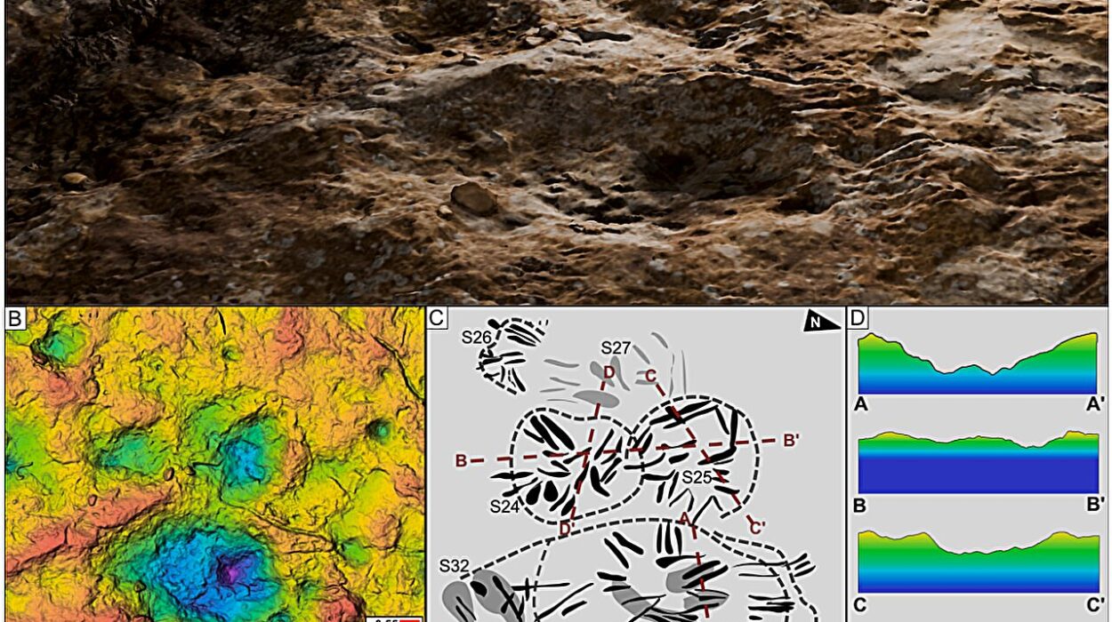
The fossil, charmingly dubbed Juracanthocephalus (Latin for “Jurassic thorny head”), was pulled from the Daohugou fossil beds, a treasure trove of Middle to Late Jurassic life. Using high-resolution scanning electron microscopy (SEM) and energy-dispersive spectroscopy (EDS), researchers examined the fossil in unprecedented detail.

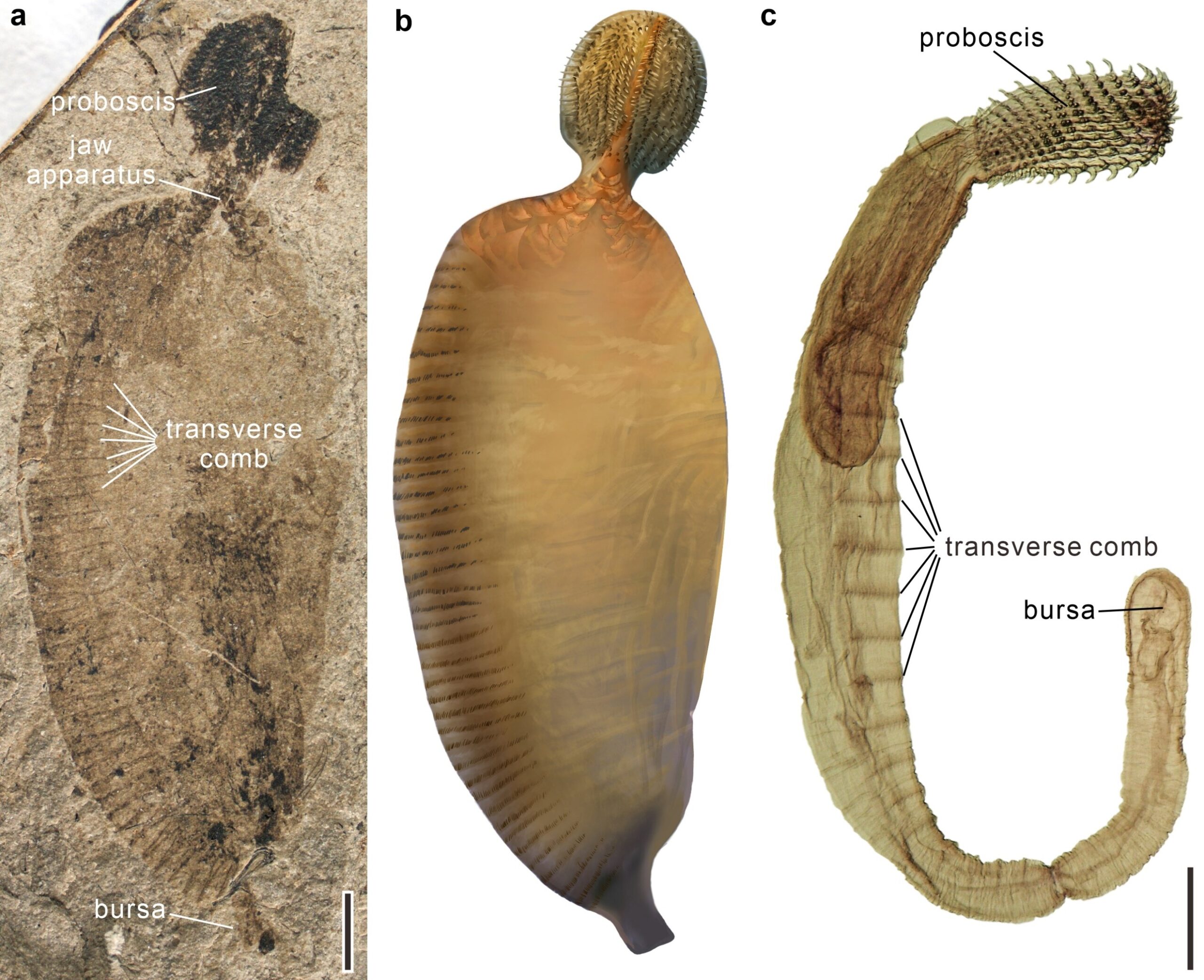
The specimen reveals a worm-like body with a division into three distinct sections: a spiny proboscis, a narrow neck, and a broader trunk. The proboscis—its most striking feature—is armed with robust, slightly curved hooks that resemble modern acanthocephalans. These hooks were likely used to anchor the parasite inside the intestines of a Jurassic-era host.
Further along its body, the researchers found 38 rows of fine, setaceous combs lining the ventral surface of the trunk—an odd but compelling morphological link to its modern relatives. These traits, combined with the presence of a jaw-like apparatus composed of tiny, tooth-like structures arranged in paired rows, link Juracanthocephalus to the broader group Gnathifera, which includes both rotifers and other jawed microfauna like Micrognathozoa and Gnathostomulida.
The jaws increase in size posteriorly and appear to be embedded in a pharyngeal framework—offering a tantalizing clue to the creature’s feeding strategy and its position on the evolutionary timeline. The posterior end of the fossil even displays a structure reminiscent of the bursa, a reproductive feature in male acanthocephalans.
Bridging the Gap: Rotifers, Parasites, and the Mystery of Morphological Mayhem
So how do you get from a free-swimming rotifer with jaws and a digestive tract to a gutless, parasitic acanthocephalan? For decades, evolutionary biologists have debated this question. The shift involves not just a new lifestyle—endoparasitism—but radical changes to the body plan. The transition seemed too dramatic, the morphological leap too vast.
But Juracanthocephalus is the long-missing intermediate. In a revised morphological matrix comparing modern and fossil worm-like animals, this fossil was positioned as a stem-group acanthocephalan—a relative sitting just outside the crown group of modern thorny-headed worms.

And here’s where it gets even more interesting: when Juracanthocephalus was excluded from the phylogenetic analysis, morphological data alone supported one arrangement—placing Seisonidea (a rare rotifer group) as the sister group to all other rotifers. But when Juracanthocephalus was included, the analysis shifted. Now, Seisonidea appeared as sister to Juracanthocephalus and modern acanthocephalans, suggesting a more nuanced and accurate evolutionary pathway.
In other words, this single fossil helped reconcile the tension between molecular and morphological evolutionary trees—two frameworks that often diverge like squabbling siblings.
Jurassic Parasite, Terrestrial Origins?
One of the most provocative implications of the discovery is environmental: the fossil suggests that acanthocephalans may have originated not in the sea, as previously thought, but on land.
This idea challenges long-held assumptions. Modern acanthocephalans infect aquatic hosts, leading scientists to assume a marine origin. But Juracanthocephalus comes from a terrestrial fossil site and shows features indicating a parasitic lifestyle—such as its large size and spiny head.
This opens a new hypothesis: acanthocephalans may have started their evolutionary journey infecting terrestrial or semi-terrestrial hosts during the Middle Jurassic, only later expanding into aquatic ecosystems. If true, that would upend the timeline and environmental context of their emergence, sparking fresh inquiries into how parasite-host coevolution has unfolded across Earth’s history.
Why This Discovery Matters—Way Beyond Worms
At first glance, it might seem niche: a fossil worm with a spiny head found in some Jurassic rocks. But Juracanthocephalus is a landmark discovery in evolutionary biology.
First, it provides direct fossil evidence of a lineage we’ve only been able to study through genetics and inference. For evolutionary theorists, it’s like finding a missing sentence in a long-erased chapter of the book of life.
Second, it’s a case study in how dramatic body plan changes occur. The transition from free-living rotifer to gutless parasite isn’t just weird—it’s radical. Understanding that shift helps scientists grasp how parasites evolve, how specialization happens, and how complexity can arise from simplicity (or vice versa).
Third, it demonstrates the power of fossil evidence in complementing and correcting molecular data. While DNA has revolutionized biology, fossils like Juracanthocephalus remind us that bones, hooks, and combs still have much to say.
Finally, this discovery underscores the enduring importance of places like the Daohugou Biota—Jurassic fossil beds that continue to yield stunning, perfectly preserved specimens capable of transforming scientific paradigms.
The Road Ahead
Of course, Juracanthocephalus is just one piece of a much larger puzzle. It raises as many questions as it answers. What host did it parasitize? How widespread were its relatives? Did similar transitional forms exist in marine settings? Can we find more fossils that track the evolutionary shift from jawed to jawless, gut-bearing to gutless, free-living to parasitic?
The answers lie buried in ancient rocks, waiting for sharp eyes and sharper tools. But for now, this tiny fossilized worm is making a giant splash in evolutionary biology, transforming how we see some of nature’s most bizarre and beguiling creatures.
Reference: Bo Wang, A Jurassic acanthocephalan illuminates the origin of thorny-headed worms, Nature (2025). DOI: 10.1038/s41586-025-08830-5. www.nature.com/articles/s41586-025-08830-5