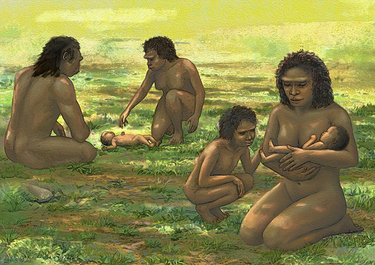
Long after their owners had died, a handful of fossil skulls preserved an unexpected kind of asymmetry that is familiar to many modern pediatricians. By tracing those subtle distortions through fossils of Homo erectus and Homo floresiensis, researchers argue that some ancient members of our genus may have begun life much like modern human babies—born unusually helpless, with soft, flexible skulls that depended on prolonged care during their earliest months.
Modern human babies arrive in the world in a remarkably unfinished state. They cannot hold up their heads, crawl, or walk for months. Their skull bones remain thin and only loosely connected, allowing the brain to continue an extraordinary burst of growth after birth. This extended period of dependence demands years of intensive care from parents and often from other members of a social group as well.
Exactly when this distinctive stage of human life evolved has remained one of paleoanthropology’s longest-running questions. Fossils preserve bones, not behavior, making it difficult to determine whether extinct human relatives experienced the same prolonged helpless infancy seen today.
Instead of relying on indirect clues such as brain size or pelvic anatomy, the new study pursued an entirely different line of evidence—one hiding in the shape of ancient skulls themselves.
A childhood condition became an unexpected window into deep evolutionary history
The researchers centered their investigation on deformational plagiocephaly, a form of skull asymmetry that develops in many healthy modern human infants.
This condition usually appears during the first months after birth when several circumstances come together. Human newborns spend long periods lying on flat surfaces, their neck muscles are still too weak to consistently move their heads, and their skull bones remain soft and flexible while the rapidly growing brain pushes outward from within.
Pressure on one part of the skull can gradually flatten that area and produce a distinctive asymmetrical shape. Clinical studies have shown that these deformities become most noticeable during the first four to six months of life, often peaking while infants still have limited head control. As babies grow stronger, many of these distortions improve, although pronounced cases can remain throughout life.
The researchers argue that this process depends on a unique combination of biological features that modern humans possess. Other living great apes develop differently. Their skull bones mature more quickly, newborns gain head control much earlier, and infants cling directly to their mothers instead of lying independently on flat sleeping surfaces. Together, those differences should make pronounced deformational plagiocephaly extremely uncommon outside humans.
If that assumption is correct, unusually large skull asymmetries in fossil humans could preserve direct evidence that ancient infants also experienced prolonged helplessness.
Looking for a human signature among more than 1,500 skulls
To test this idea, the team assembled an unusually large comparative dataset.
They examined 1,504 skulls in total, including 508 modern human specimens and 996 great ape skulls representing chimpanzees, bonobos, gorillas, and orangutans. The human sample combined CT scans from 123 healthy Japanese infants between 27 and 545 days old with hundreds of well-preserved historical skulls from Japan. The great ape collection included individuals spanning different ages and both sexes, excluding only some robust adult males whose heavy cranial crests complicated the measurements.
The infant CT scans also allowed the researchers to document how the human skull changes during the first year and a half of life. Open fontanelles—the familiar “soft spots”—and connecting sutures gradually closed as infants matured, while the greatest skull asymmetries tended to appear roughly 50 to 150 days after birth before decreasing as babies developed better control of their heads.
The researchers then measured skull asymmetry using two different directions.
One measured differences running diagonally across the skull from front to back, capturing the type of flattening commonly associated with positional plagiocephaly. The other measured side-to-side tilting across the skull. By comparing both measurements across humans and great apes, they could determine whether fossil skulls fell within the normal variation seen in living apes or exceeded it.
Modern humans stood apart from every great ape examined
The comparison revealed a clear pattern.
All four great ape groups displayed some natural asymmetry, regardless of age or sex. However, those differences remained relatively modest.
Modern humans were different.
Across both measurement methods, human skulls showed substantially greater variation than the pooled great ape sample. Some people exhibited skull asymmetries far beyond anything seen in the living apes, matching the expectation that pronounced deformational plagiocephaly reflects the unusually prolonged helpless infancy unique to humans.
The researchers also explored whether ordinary developmental asymmetry, brain lateralization, or differences in skull shape among species could explain the results. They concluded that these factors were unlikely to account for the large gap separating humans from great apes.
That provided a benchmark for evaluating fossil members of the genus Homo.
Three ancient skulls crossed a boundary rarely seen outside modern humans
The fossil analysis focused on six exceptionally well-preserved adult skulls from Indonesia.
Five belonged to Homo erectus, while one was LB1, the famous type specimen of Homo floresiensis, the diminutive species discovered on the island of Flores.
Three specimens immediately attracted attention.
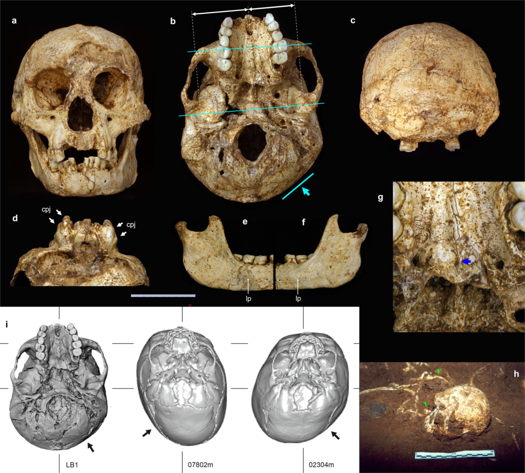
LB1 possessed horizontal skull asymmetry that exceeded every great ape in the dataset. Its measured difference reached 7.8 millimeters, compared with a maximum of 5 millimeters among the apes, while its relative asymmetry reached 5.7%, again exceeding the ape maximum of 3.7%.
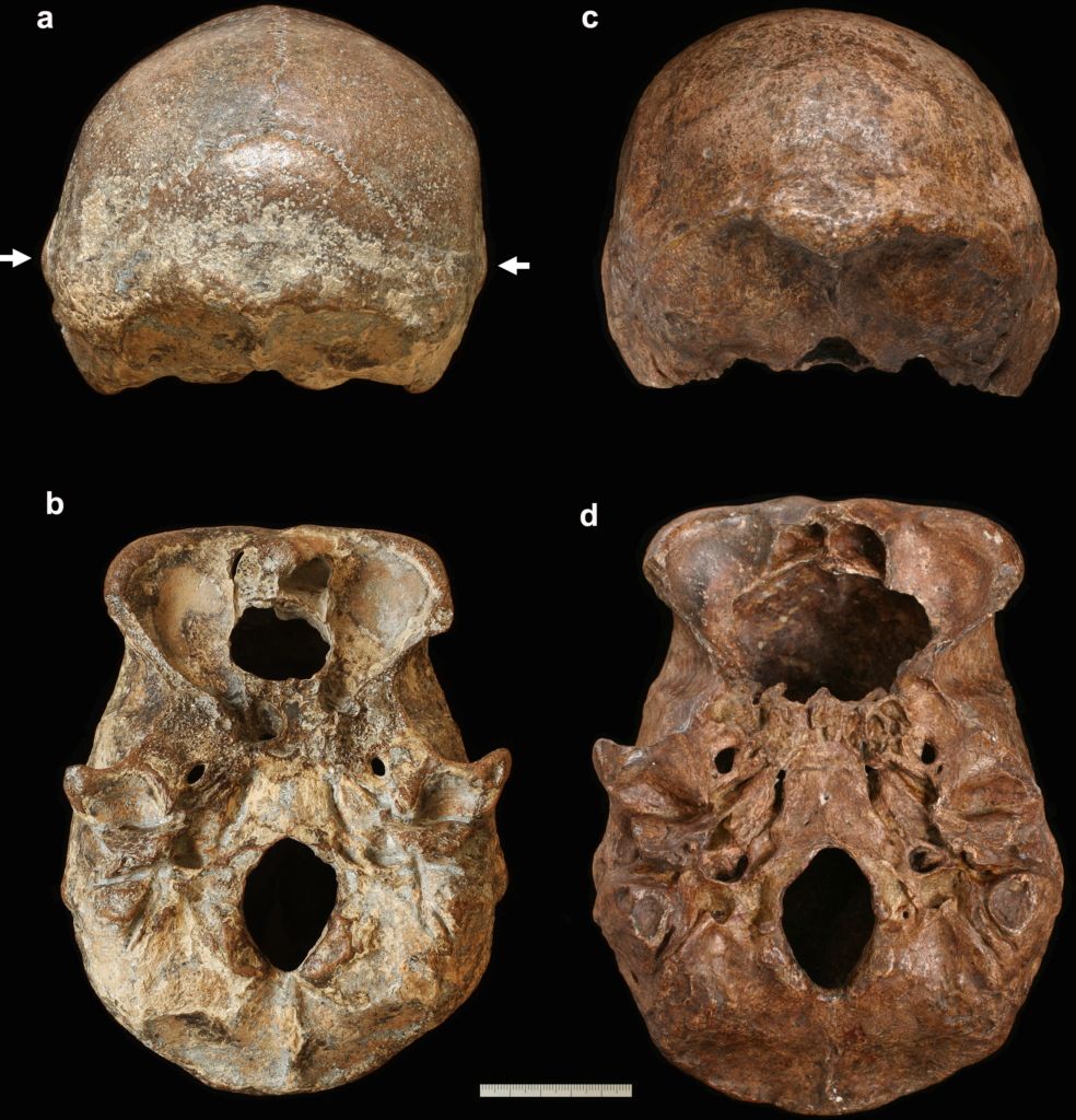
Two Homo erectus skulls—Ngawi 1 and Ngandong 12—showed unusually large side-to-side skewing. Their raw measurements reached roughly 6.7 to 6.9 millimeters, beyond the greatest value recorded among the great apes.
Although two ape specimens displayed similar percentage values for one measurement, only 0.2% of the ape sample reached that level. The authors calculated that randomly selecting two such highly distorted skulls among five ape specimens would have a probability of only 0.004%.
When both kinds of asymmetry were considered together, all three fossil skulls fell outside the estimated range of variation for the great apes.
The researchers asked whether the distortions happened after death
Finding an unusual skull shape is only part of the story.
Fossils can become distorted while buried underground, compressed by sediments or damaged during fossilization. To support their interpretation, the researchers closely examined whether these asymmetries instead reflected life before death.
For LB1, they analyzed the original fossil, high-resolution CT scans, and excavation records.
The skull had been discovered remarkably well preserved, along with other fragile skeletal bones, in fine cave sediments deposited under calm conditions. Internal anatomical features remained aligned, and the asymmetry extended across multiple facial structures rather than appearing as isolated damage. The researchers also noted that the direction of burial pressure could not explain the observed pattern of twisting.
The two Homo erectus skulls presented similar evidence.
Although both lacked facial bones, the remaining crania retained smooth bone continuity without the fractures or internal disruptions expected from major postmortem distortion. CT images showed robust skull regions displaying pronounced asymmetry while delicate structures remained largely undisturbed, a pattern the authors considered inconsistent with burial damage.
Taken together, they concluded that the most likely explanation was that these individuals had experienced pronounced deformational plagiocephaly during infancy.
The authors acknowledge that some contribution from postmortem forces can never be ruled out completely, but they argue that such effects were unlikely to be the primary source of the observed skull shapes.
What these ancient infants may have been like
If the interpretation is correct, the implications extend far beyond skull anatomy.
Pronounced deformational plagiocephaly develops because infants possess soft, rapidly growing skulls, weak neck muscles, and spend considerable time lying with their heads supported. Those characteristics collectively describe the prolonged helpless infancy that distinguishes modern humans.
The findings therefore suggest that at least some populations of Indonesian Homo erectus and Homo floresiensis shared this developmental pattern despite having considerably smaller brains than modern humans.
That idea is particularly striking for Homo floresiensis.
LB1’s brain volume measured 426 cubic centimeters, comparable to estimates for Australopithecus afarensis, while its body size also overlapped with that much earlier hominin. Yet the apparent presence of deformational plagiocephaly implies that its early brain and skull development differed from Australopithecus despite the similar adult brain size.
Rather than representing a simple return to an earlier developmental pattern as brain size decreased, Homo floresiensis may have retained a distinctly human style of infancy while evolving smaller brains on its isolated island.
Caring for helpless infants may have become part of Homo life much earlier than expected
The study also carries behavioral implications.
If infants of Homo erectus and Homo floresiensis were as physically dependent as the skull evidence suggests, adults would have faced many of the same challenges associated with caring for helpless babies today.
The authors note that Indonesian Homo erectus lived alongside predators including tigers and leopards, while Homo floresiensis shared its island with Komodo monitors and giant carrion birds. Successfully raising vulnerable infants under those conditions would have required effective protection, sustained parental care, and likely cooperation within social groups.
The researchers emphasize that this interpretation remains an inference built upon developmental evidence rather than direct observation of behavior. Even so, the skulls hint that elaborate caregiving may have become established across multiple branches of the genus Homo.
The fossils open a new path, but many questions remain
The researchers see this work as a starting point rather than a final answer.
Their approach can only be applied to fossils that are exceptionally well preserved and free from significant postmortem distortion. Many older hominin skulls are too incomplete for the same analysis.
Whether helpless infancy first evolved once in a common ancestor or appeared independently in different human lineages also remains unresolved. The presence of similar skull deformities in Neanderthals, together with the new evidence from Indonesian Homo erectus and Homo floresiensis, raises the possibility that this developmental strategy originated deep in the evolutionary history of Homo. However, the authors stress that older fossil material must be examined before that question can be answered.
For now, the study suggests that subtle asymmetries preserved in ancient skulls may record something unexpectedly intimate: not how these individuals died, but how they began life. By recognizing the lasting imprint left by infancy, researchers may have uncovered a new way to trace the emergence of one of humanity’s defining characteristics—the unusually long, vulnerable childhood that made prolonged brain growth, intensive caregiving, and perhaps new forms of social life possible.
Publication details
Kaifu Y, et al. Cranial evidence for human-like helpless infancy in Homo erectus and Homo floresiensis, Proceedings of the Royal Society B: Biological Sciences (2026). DOI: 10.1098/rspb.2026.1055