For as long as humans have peered through lenses, the microscope has been our gateway to hidden worlds. From Robert Hooke’s first drawings of cork cells to the dazzling fluorescent images of living cells today, microscopes have extended our vision into realms once unimaginable. Yet, biology is full of processes so small, so fast, and so delicate that even our most powerful tools struggle to capture them without disturbing the very life we seek to observe.
Super-resolution microscopy, developed in the past two decades, has shattered the so-called diffraction limit of light, allowing us to see structures far smaller than what classical optics would permit. But in live-cell imaging—where the goal is not just to see but to watch life unfold in real time—scientists face a painful tradeoff. To gain sharper detail, they often sacrifice speed, and in trying to observe longer, they risk damaging the very cells they hope to study. This is where the breakthrough from Professor Xi Peng’s team at Peking University enters the story.
The Promise and Problem of SIM
Among super-resolution techniques, Structured Illumination Microscopy (SIM) has stood out as the gentlest option for live-cell imaging. Unlike harsher methods that flood cells with light, SIM uses patterned illumination to extract fine structural details. By shining stripes of light at different angles and mathematically reconstructing the images, SIM doubles resolution while keeping cells alive and responsive.
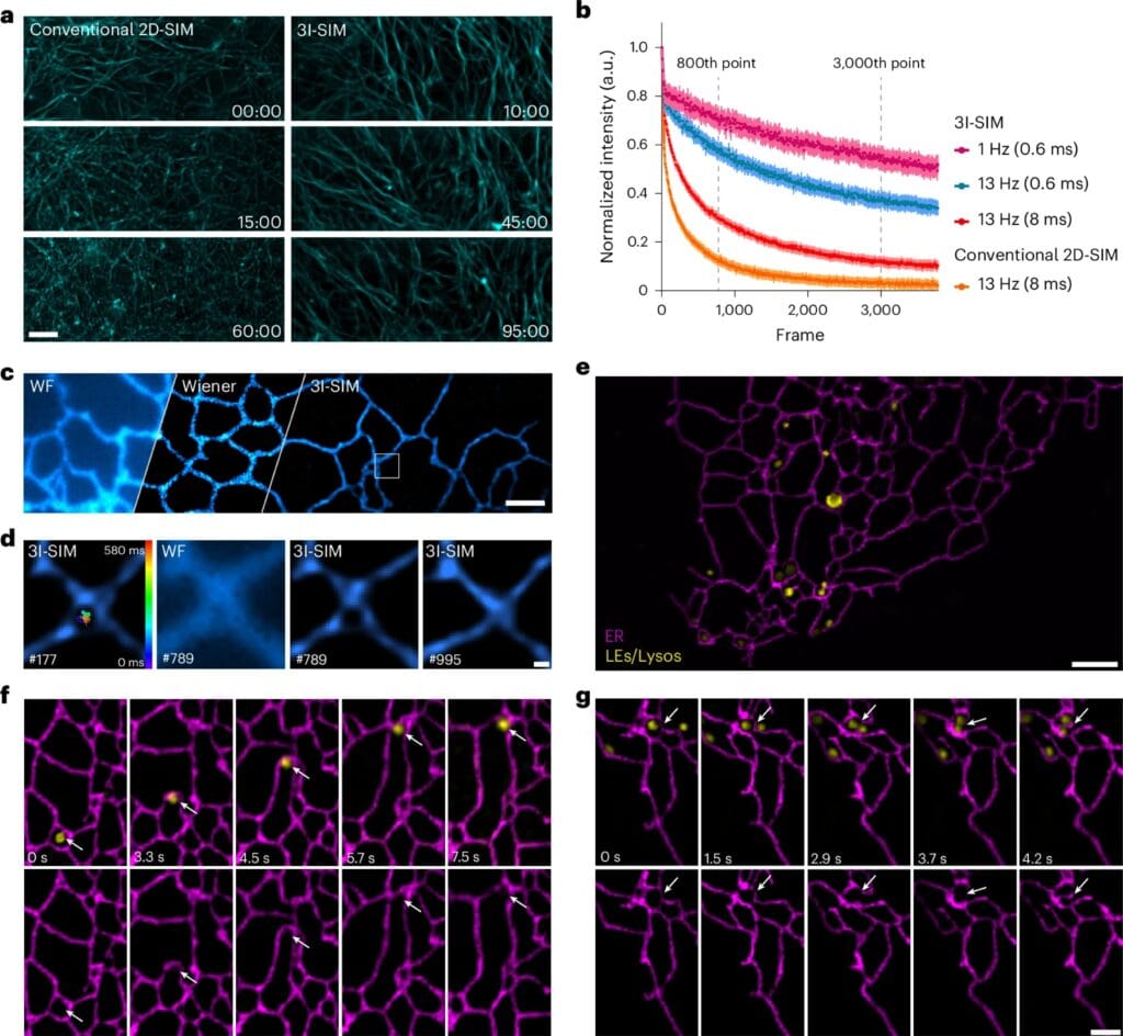
But conventional SIM comes with a hidden cost. To build a uniform high-resolution image, the system requires multiple exposures—typically nine separate images for each frame—because the striped illumination must be rotated in three different directions. This slows down imaging speed, consumes precious photons, and inevitably causes photobleaching, a fading of fluorescence that signals damage to cells. As a result, researchers are left watching only fragments of cellular life, unable to follow its most rapid and intricate dances.
A New Shape of Light
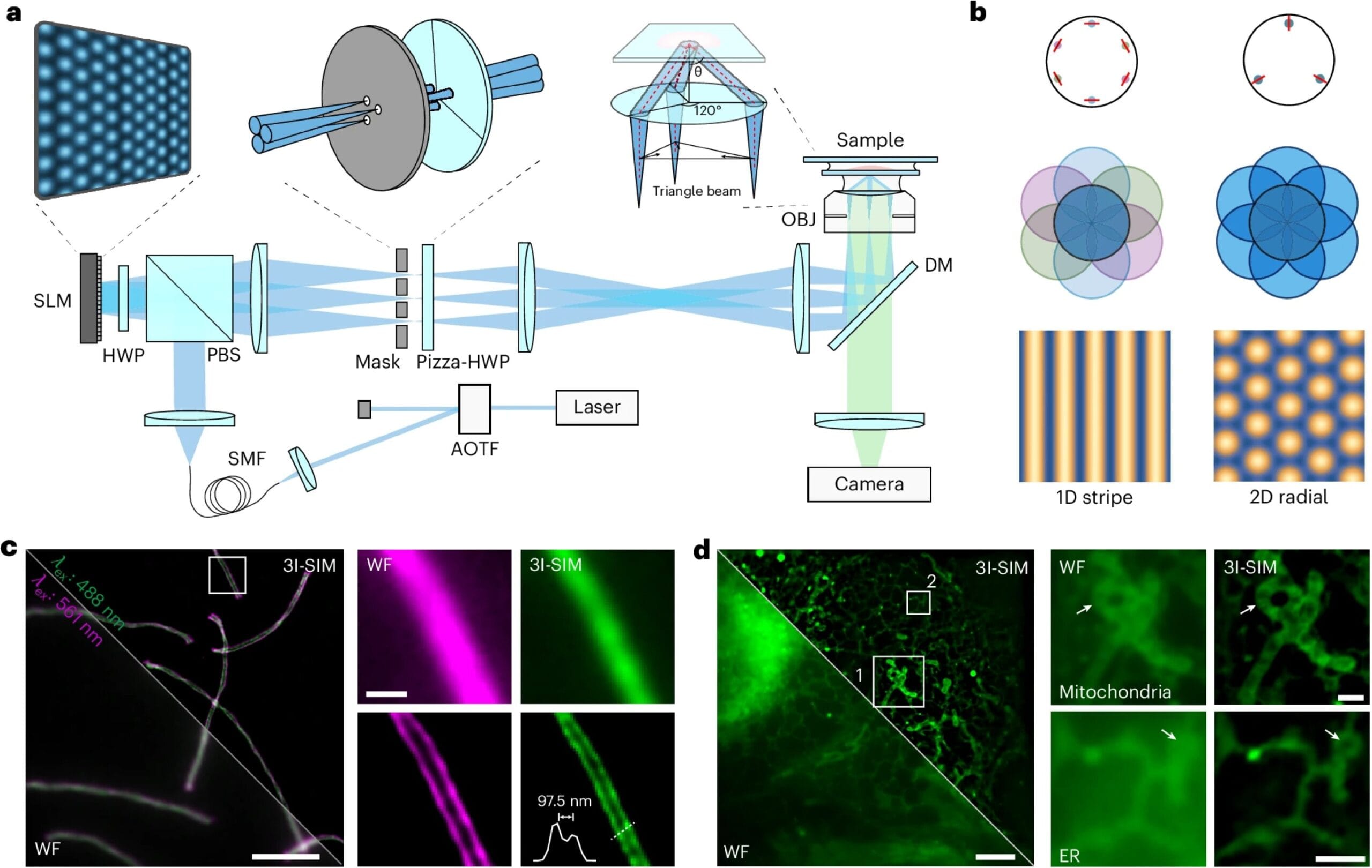
Professor Xi Peng and his colleagues asked a deceptively simple question: what if illumination could be patterned not by rotating stripes, but by weaving beams of light together in a more efficient geometry? Inspired by the strength and elegance of triangular structures, they developed Triangle-Beam Interference SIM (3I-SIM)—a method that replaces rotating stripes with a stable hexagonal lattice of light.
Instead of the laborious nine exposures required in classical SIM, 3I-SIM reconstructs images with just seven frames, and these frames are shifted along only one direction. This innovation slashes redundancy and unlocks breathtaking speed. The system achieves imaging at up to 1,697 frames per second, a rate fast enough to capture fleeting cellular events once considered unobservable. Even more striking, the gentler illumination means that imaging can continue for up to 13 hours straight, yielding over 100,000 snapshots of life in action.
Capturing Life in Motion
The power of 3I-SIM is not in numbers alone but in the new biology it makes visible. With this tool, scientists recorded delicate shape changes in neuronal growth cones, the dynamic tips of neurons that explore their environment and guide brain wiring. They also tracked transient actin signals that control the movement of the endoplasmic reticulum, one of the cell’s busiest organelles. These fleeting events, which flicker and vanish in moments, had long eluded researchers. Now, with sustained, high-speed imaging, they can be followed in exquisite detail over hours of living activity.
For cell biology, this is akin to switching from a handful of blurry photographs to a full-length documentary film. Processes that once appeared as static snapshots can now be understood as dynamic stories, unfolding second by second within the living cell.
Lowering Barriers, Expanding Horizons
One of the most remarkable aspects of 3I-SIM is its accessibility. Unlike some advanced imaging systems that require entirely new instruments, the triangle-beam method can be upgraded onto existing 2D-SIM platforms. This means research labs around the world, even those without massive budgets, can adopt the technology and begin exploring the fast and fragile processes that define cellular life. By reducing technical barriers, the innovation opens the door for a broader community of scientists to contribute discoveries.
A Leap Forward for Biology
The publication of this research in Nature Photonics signals more than a technical advance. It represents a leap forward in how we can study life at its smallest scales. For decades, researchers have dreamed of watching cells work in real time without harming them, capturing the beauty of living systems as they are, not as frozen or distorted artifacts. With 3I-SIM, that dream comes closer to reality.
Einstein once said that the most beautiful experience we can have is the mysterious. For modern biologists, the mysteries lie not in distant galaxies but within the bustling world of a single cell. Thanks to innovations like 3I-SIM, we are now equipped to explore those mysteries with clarity, gentleness, and speed, bringing us closer to understanding the vibrant and dynamic essence of life itself.
More information: Yunzhe Fu et al, Triangle-beam interference structured illumination microscopy, Nature Photonics (2025). DOI: 10.1038/s41566-025-01730-0