“The light of someone’s life” is often a poetic metaphor. But what if that light is literal? According to groundbreaking research from the University of Calgary, all living systems—plants, animals, even humans—emit a ghostly, ultraweak light invisible to the naked eye. This phenomenon, known as Ultraweak Photon Emission (UPE), may serve as a biological fingerprint of life itself.
In an era where health diagnostics are becoming increasingly non-invasive and sensitive, the ability to detect light that living organisms naturally emit—without any external excitation—opens a new frontier. A quiet glow, not seen but sensed through scientific instrumentation, might soon offer insights into vitality, stress, and disease.
Biophotons: The Subtle Light of Life
Unlike the dazzling spectacle of fireflies or deep-sea creatures using bioluminescence, UPE is extraordinarily dim—millions of times fainter than what our eyes can perceive. This emission, often dubbed biophoton emission, occurs across the ultraviolet to near-infrared spectrum (200–1,000 nm). It’s not just a biological curiosity; it may be a vital sign of life itself.
UPE doesn’t require energy input from an external light source, like fluorescence does. Instead, it arises from internal biochemical processes, particularly those associated with oxidative metabolism—the essential machinery of life that turns food, oxygen, and sunlight into cellular energy.
Living Chemistry and the Glow Within
To understand UPE, we must step into the dynamic microcosm inside every living cell. Here, metabolic reactions are constantly producing reactive oxygen species (ROS)—molecules that contain oxygen and are highly reactive due to unpaired electrons. These ROS are natural byproducts of oxygen metabolism and play crucial roles in cell signaling, immune responses, and even development.
However, when ROS levels exceed the cell’s antioxidant defenses, oxidative stress ensues. This can damage proteins, lipids, and DNA. But more subtly, it can also excite electrons in biomolecules. As these electrons return to their ground state, they release tiny flashes of light—UPE.
This is not mere background noise. UPE reflects the redox (oxidation-reduction) status of the cell, which makes it a sensitive reporter of metabolic activity, stress, injury, and potentially disease.
Seeing the Invisible: How Scientists Measure UPE
To detect this faint light, scientists must first eliminate all other sources of illumination. The researchers at the University of Calgary constructed ultradark enclosures—light-sealed boxes akin to sensory deprivation chambers for photons. Within these, they placed either live mice or plants and observed them using ultra-sensitive cameras.
For imaging animals, the team used a Charge-Coupled Device (CCD) camera integrated with the IVIS system, which is typically used in preclinical imaging. For plants, they employed an Electron-Multiplying CCD (EMCCD) camera, which can detect even the weakest light signals.
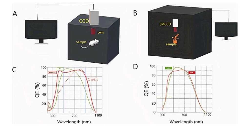
Both systems were calibrated to pick up emissions in total darkness, ensuring that any detected light came solely from the organism itself.
The Glow of Life and the Silence of Death
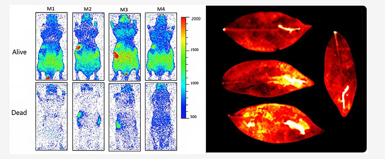
One of the most striking findings came from comparing UPE in living versus recently euthanized mice. Despite both sets of animals having the same body temperature—37°C, a key control to eliminate thermal interference—only the live mice emitted measurable levels of UPE.
Their glow was consistent and unmistakable. In contrast, the bodies of dead mice fell into a photon silence, their metabolic machinery stilled. This sharp contrast suggests that UPE is closely tied to active life processes rather than mere warmth or residual activity.
In effect, UPE could be described as a “vital light,” an ephemeral signature that fades not with the body’s temperature, but with the extinguishing of cellular function.
Plants Under Pressure: Stress Glows Bright
Plants, too, emitted UPE, but their glow responded dynamically to environmental stress. The researchers subjected Arabidopsis thaliana, a model plant species, to various stressors including mechanical injury, temperature changes, and chemical treatments.
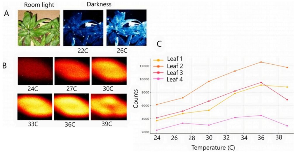
When a leaf was wounded, the damaged area lit up significantly brighter than surrounding healthy tissue. Exposure to heat also triggered a marked increase in photon emission. The addition of hydrogen peroxide, a known ROS stimulant, caused another spike. Conversely, when antioxidants were applied, UPE levels dropped, suggesting a direct correlation with oxidative stress.
Interestingly, even uninjured areas began to emit more light after the injury, hinting at a systemic stress response—a possible communication signal between different parts of the plant. This observation opens up new avenues to study plant behavior and resilience using UPE as a non-invasive biomarker.
What’s Behind the Glow? ROS and Energy Transitions
So, why exactly does stress make living systems glow?
The answer lies in the electron transitions within biomolecules, especially lipids and proteins. Under oxidative stress, molecules like singlet oxygen and excited carbonyls are formed. As these high-energy molecules return to their ground state, they release photons.
These photons aren’t coordinated or intentional like those in bioluminescence. They are spontaneous, probabilistic releases—the molecular equivalents of sighs from overworked systems. Yet, their patterns carry biological meaning. They may help researchers decode the invisible signatures of stress, injury, and vitality.
A Tool for the Future: From Diagnostics to Biophysics
The implications of this research stretch far beyond curiosity.
UPE imaging could become a powerful tool in non-invasive diagnostics. Imagine detecting early oxidative stress in human tissues without drawing blood or exposing patients to radiation. Or monitoring plant health in agriculture without cutting or sampling. The technique could even track the progression of diseases like cancer, neurodegeneration, or infections—all of which are associated with metabolic changes and oxidative stress.
In neuroscience, for instance, researchers are beginning to explore whether UPE can detect subtle metabolic shifts in the brain—potentially revealing early signs of disorders before symptoms appear. In regenerative medicine, it might help track tissue repair in real time.
From a pure science perspective, UPE offers a way to study life at its most fundamental level—how energy flows through living systems, how stress alters that flow, and how resilience is encoded in light.
Light as the Language of Life
One of the most poetic aspects of UPE is that it repositions light not merely as something we use to see the world, but something life uses to signal its own internal state. It suggests that life is not only powered by light, as in photosynthesis or vision, but also quietly speaks through it.
This bridges ancient intuitions and modern science. For millennia, mystics and healers spoke of “auras” or inner light. While not mystical in itself, UPE research shows that biological systems do, indeed, radiate a form of internal light—one that could inform science, medicine, and our broader understanding of life.
Challenges and Future Horizons
Despite its promise, UPE imaging is still in its infancy. The signals are faint and can be affected by numerous factors—temperature, oxygen availability, biological variability. Instrumentation must be highly sensitive and environments carefully controlled.
Moreover, while UPE tells us that stress is occurring, decoding exactly why or where in the body the stress originates is still a complex challenge. Integrating UPE imaging with other technologies like spectroscopy, machine learning, and molecular probes may help refine its diagnostic power.
Nevertheless, the fact that we can see life glow—even faintly—is extraordinary. It offers a new dimension to the way we monitor health, study resilience, and understand mortality.
Conclusion: A New Light on Life Itself
In the end, the University of Calgary study does more than reveal a novel biological phenomenon. It challenges us to reconsider the boundaries between biology, physics, and even philosophy.
Light, often seen as a metaphor for consciousness, inspiration, or spirit, may also be a literal byproduct of being alive. The cells in your body, the leaves on a tree, even single bacteria in the soil—they all hum with an unseen shimmer, a soft whisper of photons telling the story of their inner worlds.
This gentle, invisible glow may soon become a powerful lens through which we detect stress, diagnose illness, and monitor vitality. It reminds us that even in darkness, life glows on.
Reference: V. Salari et al, Imaging Ultraweak Photon Emission from Living and Dead Mice and from Plants under Stress, The Journal of Physical Chemistry Letters (2025). DOI: 10.1021/acs.jpclett.4c03546