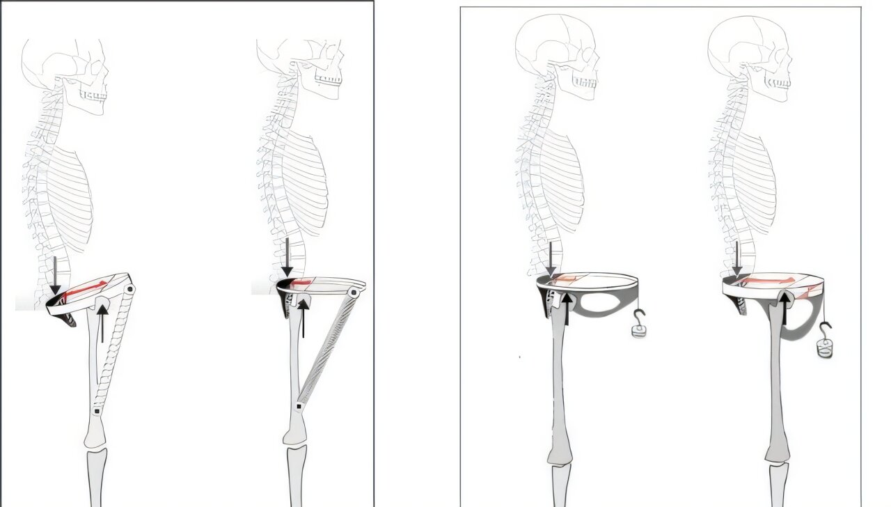
For decades, the Neanderthal pelvis has been treated as the unusual one. A comparison of Neanderthal and modern human pelvises now points in the opposite direction: the distinctive anatomy may be the modern human male pelvis, whose hip joints are positioned in a way that creates a different mechanical system for walking.
The comparison that changed the picture
The study compared two nearly complete male Neanderthal pelvises with dozens of modern human pelvises. One Neanderthal pelvis came from Kebara Cave in Israel, while the other was found at Sima de los Huesos in Spain.
Despite being large and robust, the Neanderthal pelvises matched modern human female pelvises in most of the measurements and proportions examined. They did not show the distinctive configuration seen in modern human males.
That finding led the researchers to reconsider which pelvis should be regarded as unusual.
The ancestral pelvic configuration may have been retained in Neanderthals and modern human females, while the modern human male pelvis underwent substantial evolutionary changes.
The position of the hip joints is central
The most important difference concerns the position of the hip joints.
In modern human males, the hip joints are located farther forward on the pelvic ring than they are in modern human females and male Neanderthals. According to the researchers’ biomechanical model, this change in position created a different way for the pelvis and thigh muscles to handle the forces produced during walking.
The muscles at the front of the thigh attach to the front of the pelvis. With the altered geometry of the male pelvis, the researchers propose that these muscles can act somewhat like a spring, while body weight loads the back part of the pelvis.
This arrangement provides the basis for the study’s proposed shock-absorbing and energy-return mechanism.
How the proposed mechanism works during walking
During each step of bipedal walking, the body’s center of mass moves downward. That downward movement places stress on the joints and requires energy to raise the body again for the following step.
The researchers propose that the distinctive geometry of the modern human male pelvis allows the thigh muscles to cushion this downward movement. As the body drops, the system can store potential energy and then release it immediately afterward.
In the model, that stored energy helps propel the body upward into the next step.
The researchers therefore describe the pelvis as functioning as a natural shock absorber and energy-return system. They propose that this mechanism can reduce energy expenditure and make walking more efficient, potentially providing an advantage during long-distance travel on foot.
The altered hip position also came with other structural changes. The researchers identify a thicker pubic bone and a deeper anterior part of the pelvis as adaptations needed to withstand the changed mechanical loads.
Why the female pelvis remained different
The researchers argue that modern human females could not adopt the complete set of these modifications.
Childbirth places constraints on pelvic structure, requiring a relatively shallow pelvis and a birth canal that is sufficiently wide. According to the study’s interpretation, these constraints left the modern human female pelvis closer to the ancestral configuration.
That configuration is also found in the male Neanderthal pelvises examined in the study.
This provides the basis for the researchers’ explanation of part of the sexual dimorphism in the human pelvis, meaning the anatomical differences between males and females.
The evolutionary change may have occurred in modern human males
The comparison changes the evolutionary framing of the pelvis.
Rather than treating the Neanderthal pelvis as an unusual structure that needs a special explanation, the researchers argue that the distinctive anatomy is the modern human male pelvis. In their model, the male configuration represents the evolutionary innovation, while Neanderthals and modern human females retained the more ancestral arrangement.
Professor Ella Been, a co-author of the study, said the findings could also contribute to research involving biomechanics, musculoskeletal medicine, rehabilitation and injury prevention.
The researchers present their work as a new biomechanical model for understanding a substantial part of the anatomical differences between male and female human pelvises. They also argue that the findings demonstrate that previously unrecognized structures, geometries and biological mechanisms can still be identified in human macroscopic anatomy.
The study was published in Scientific Reports.