It’s a small miracle we rarely think about. Every second, countless light signals hit our eyes from different directions, at different distances, and along wildly different neural pathways. These signals should arrive at the brain at slightly different times — yet our vision feels seamless, fluid, and perfectly in sync. How does that happen?
A new study from the Institute of Molecular and Clinical Ophthalmology Basel (IOB), the University of Basel, and ETH Zurich, published in Nature Neuroscience, has revealed a hidden mechanism in the human eye that keeps visual information synchronized before it even reaches the brain. And it all happens inside the retina — the very first stage of vision.
The Eye’s First Job: Making Sense of Light
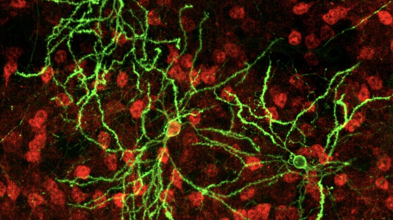
Every moment we’re awake, light pours into our eyes. At the back of each eye lies the retina — a thin sheet of neural tissue packed with millions of photoreceptors. These specialized cells capture light and turn it into tiny electrical pulses that travel along retinal ganglion cells toward the brain.
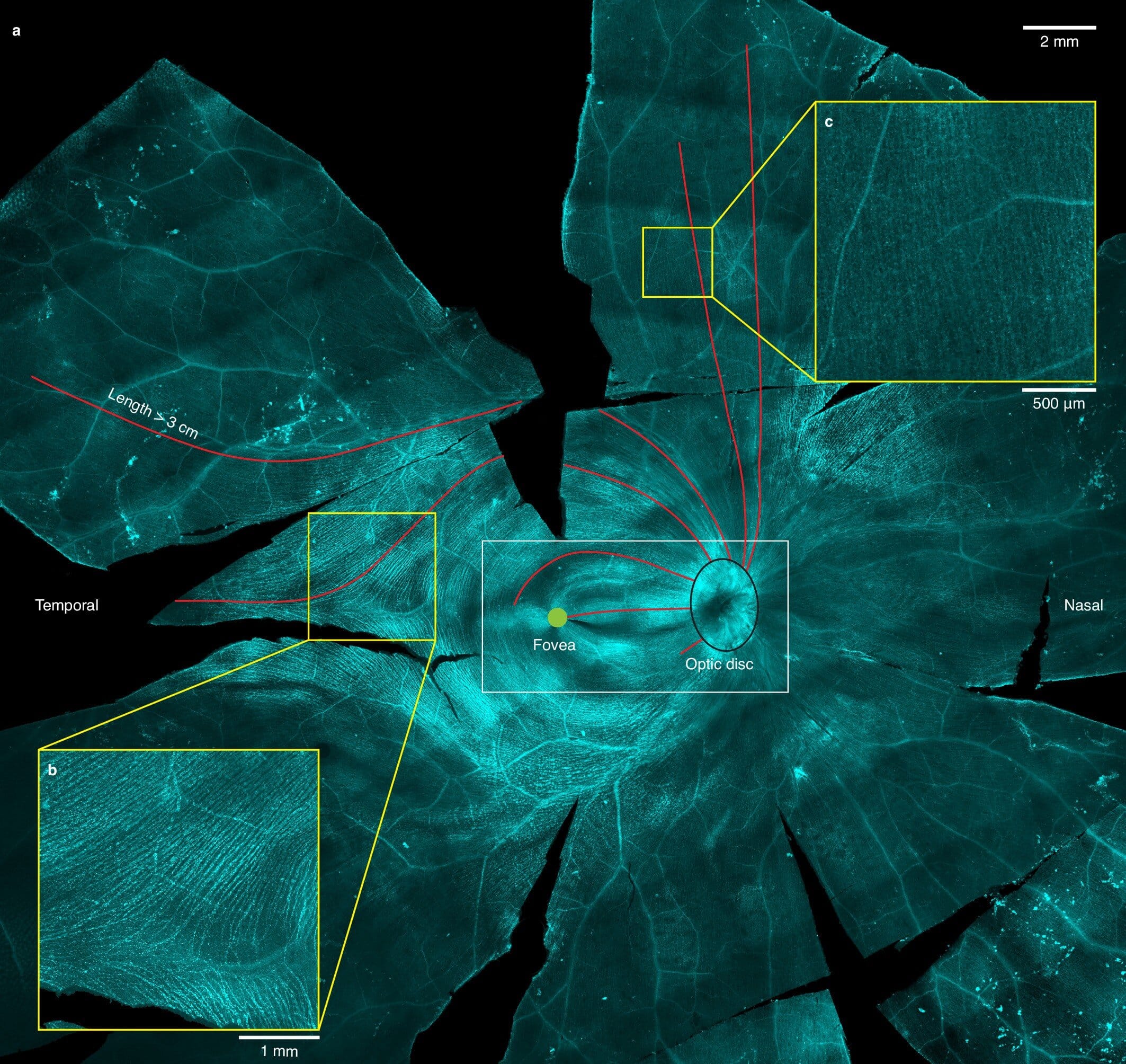
But not all parts of our vision work the same way. In the very center of the retina lies the fovea, a small pit responsible for our sharpest vision — the place we use to read words, recognize faces, and spot fine details. This region has no tolerance for distortion. In fact, axons — the long, cable-like extensions of ganglion cells — are not allowed to cross over the fovea because their presence would blur our central vision. Instead, they must bend around it, taking longer and more complex routes to the brain.
That detour creates a challenge: visual signals traveling on longer axons should take more time to arrive. If the brain received them at different moments, our perception could feel jittery and out of sync.
Yet it doesn’t. The question is — why?
A Simple Observation That Sparked a Big Question
Lead researchers Felix Franke and Annalisa Bucci began with an anatomical curiosity: the retina’s axons follow different paths depending on where they originate, and some of those paths are much longer than others. If signal speed is constant, the longer routes should produce delays.
“Our goal,” Franke explained, “was to find out whether the retina itself helps coordinate the timing of visual signals before they even reach the brain.”
That question touches on one of neuroscience’s most intriguing puzzles: how sensory systems maintain split-second timing despite the very real physical differences in the pathways carrying their signals.
Peering into the Living Retina
To answer it, the team needed to measure exactly how fast signals traveled along different retinal axons — and to do so with incredible precision. They turned to an unusual and rare resource: whole human eyes donated for research.
In the lab, they kept the retina alive and functional, so that its neurons could still send electrical signals. Using high-density microelectrode arrays, they recorded the tiny voltage changes as signals left different parts of the fovea. These devices, sampling at 20,000 times per second, captured the timing of each signal with microsecond accuracy.
The results showed that transmission speeds weren’t uniform. Axons carrying signals from certain parts of the retina were faster than others — sometimes much faster. And intriguingly, the speed differences lined up with axon length.
Anatomy Meets Physics
To dig deeper, the team used transmission electron microscopy to measure axon diameters down to the nanometer scale. They discovered a clear pattern: longer axons were thicker. This matters because thicker axons conduct electrical signals faster — a principle well known in neuroscience.
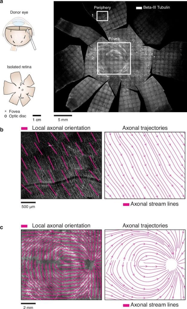
This means the retina isn’t just passively sending signals to the brain. It’s actively fine-tuning each axon’s structure so that signals from different parts of the fovea arrive at the brain at the same time, no matter how far they’ve had to travel.
“This anatomical fine-tuning is like adjusting lanes on a racetrack,” Bucci explained. “The runners on the outer lanes have more ground to cover, so we give them a head start — or, in this case, thicker axons to make them faster.”
Testing the Effect on Perception
Of course, anatomy and speed measurements are one thing — but did this synchronization actually affect what people saw?
To find out, the team used adaptive optics scanning laser ophthalmoscopy (AOSLO), a powerful imaging technique that can target individual photoreceptors in a living human eye. They flashed tiny bursts of light to single cells in different parts of the fovea and asked participants to press a button as quickly as possible when they saw the light.
If signal timing varied by location, reaction times should have varied too. But they didn’t. Across the fovea, reaction times were remarkably uniform — proof that the retina’s built-in timing adjustments were working.
A Mechanism Without Myelin
One of the most surprising aspects of this discovery is that the retina achieves this synchronization without myelin — the fatty insulation that speeds up axonal signals elsewhere in the nervous system. Myelin would block light, so the retina must remain transparent. Instead, it relies entirely on diameter adjustments to keep the signals aligned in time.
That insight could have broad implications beyond vision. “First, it tells us that unmyelinated axons can make major contributions to timing precision,” Franke said. “Second, it suggests the retina plays a far more active role in fine-tuning sensory information than we previously thought.”
Why It Matters
Precise timing in sensory systems isn’t just about image quality — it’s fundamental to how we interact with the world. A split-second delay in visual processing could throw off hand-eye coordination, make moving objects harder to track, or disrupt the delicate balance between visual and auditory perception.
The findings may also have medical implications. In diseases like glaucoma, long axons are often the first to degenerate, partly because they require more energy and are more mechanically vulnerable. Disruptions to the retina’s timing system could be an early sign of disease — or a potential target for therapy.
The team’s next steps include studying how this synchronization mechanism develops and exploring whether similar strategies exist elsewhere in the nervous system.
“Our vision feels instantaneous,” Bucci reflected. “But behind the scenes, your retina is running a perfectly tuned relay race, making sure every signal crosses the finish line together. We just discovered one of its tricks.”
More information: Annalisa Bucci et al, Synchronization of visual perception within the human fovea, Nature Neuroscience (2025). DOI: 10.1038/s41593-025-02011-3.