Imagine being able to look inside the human body without having to perform surgery, or without cutting through layers of muscle and bone. Now, imagine being able to observe organs, bones, tissues, and even the smallest fractures in real-time, from a safe and non-invasive distance. This is not the stuff of science fiction, but the powerful reality brought to us by the field of radiology.
Radiology, the branch of medicine that uses imaging techniques to diagnose and treat diseases, has revolutionized the way we understand and treat the human body. From the first x-ray taken in the late 19th century to the sophisticated imaging technologies available today, radiology has become an indispensable part of modern healthcare. Whether it’s through an x-ray, ultrasound, CT scan, or MRI, radiologists use various forms of imaging to peer inside the body in ways that were once unimaginable, enabling early detection and treatment of countless conditions.
But radiology is more than just a medical tool. It’s a scientific marvel, a field that combines technology, anatomy, physics, and medicine to create a clearer, more precise picture of human health. In this article, we will dive deep into what radiology is, how it works, the various techniques it employs, and the profound impact it has on patient care.
A Historical Overview: The Birth of Radiology
To understand radiology’s significance, it’s important to look at its roots and how it all began. The history of radiology is deeply intertwined with the discovery of x-rays, a momentous event that changed the trajectory of medical science forever.
In 1895, a German physicist named Wilhelm Conrad Roentgen made an accidental discovery that would lead to the birth of radiology. While experimenting with cathode rays, Roentgen noticed a mysterious glow emanating from a fluorescent screen in his lab, even though it was not in the direct path of the rays. Upon further investigation, Roentgen realized that this glow was caused by a new type of radiation, which he named x-rays—the “x” signifying the unknown. He soon discovered that these x-rays had the remarkable ability to pass through various materials, including the human body, while leaving behind an image on photographic plates.
Roentgen’s discovery, which he shared with the scientific community in a groundbreaking paper in 1895, marked the birth of radiology. His invention was revolutionary, as it allowed physicians for the first time to see inside the human body without surgery, providing invaluable information for diagnosis. Roentgen was awarded the first-ever Nobel Prize in Physics in 1901 for his discovery.
However, it wasn’t until the early 20th century that x-rays began to be used in medical practice. Doctors quickly realized the immense potential of this new technology. Hospitals began adopting the technology, and specialized radiological departments started to form, making radiology an essential part of healthcare.
From the invention of the x-ray, radiology continued to evolve. New technologies such as fluoroscopy, CT scans, and MRI imaging began to supplement and expand the capabilities of radiologists, leading to even greater advances in diagnosing and treating various medical conditions.
What Is Radiology?
At its core, radiology is the medical specialty that employs imaging technology to diagnose, treat, and monitor various diseases and conditions inside the human body. Radiologists are medical professionals specially trained to interpret these images, which can reveal a wide range of information about a patient’s health.
Radiology is not limited to imaging alone; it also involves the use of image-guided procedures to treat diseases. From detecting broken bones to identifying tumors, radiology plays a pivotal role in diagnosing conditions and planning treatments. Additionally, many therapeutic procedures—such as the insertion of stents, biopsies, and catheter placements—are now performed using imaging guidance, making them safer and more precise.
Radiology is often divided into two main categories:
- Diagnostic Radiology: This branch focuses on the use of imaging to diagnose medical conditions. It includes techniques like x-rays, CT scans, MRIs, ultrasounds, and fluoroscopy. Diagnostic radiologists interpret these images to identify diseases, fractures, infections, and abnormalities.
- Interventional Radiology: This is a subspecialty within radiology that involves the use of imaging techniques to guide minimally invasive surgical procedures. Interventional radiologists perform procedures such as biopsies, the removal of blood clots, and the placement of stents or catheters, all with the assistance of imaging technology. These procedures are typically less invasive than traditional surgery, resulting in shorter recovery times and fewer complications.
The Tools of the Trade: How Radiology Works
The science behind radiology lies in the interaction between energy and matter. Different imaging techniques rely on various forms of energy—whether it’s x-rays, magnetic fields, sound waves, or radiofrequency waves—to create images of the body. Each technique has its own unique properties and uses, allowing radiologists to examine different parts of the body in great detail.
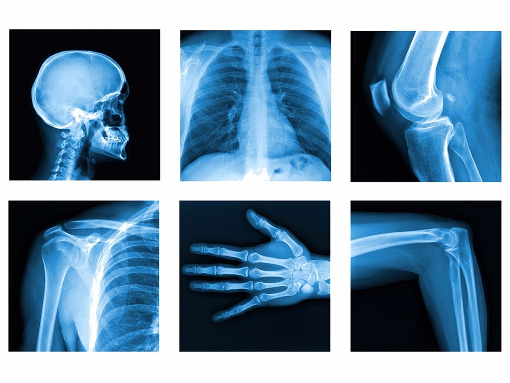
X-Ray Imaging
The classic method of radiology, x-ray imaging uses a form of electromagnetic radiation to capture images of the inside of the body. When an x-ray beam is directed at the body, different tissues absorb different amounts of radiation. Dense tissues, such as bones, absorb more x-rays and appear white on the resulting image, while softer tissues, such as muscles and organs, absorb fewer rays and appear darker. This contrast allows radiologists to identify fractures, infections, tumors, and other abnormalities.
X-rays are typically used to examine bones, joints, and the chest. They’re often the first imaging technique used when a patient presents with an injury or symptoms of a respiratory condition.
Computed Tomography (CT)
Computed tomography (CT), also known as CAT scans, is a more advanced imaging technique that uses multiple x-ray beams and a computer to create cross-sectional images of the body. These images can be combined to generate detailed 3D images, providing a much clearer and more detailed view than traditional x-rays.
CT scans are commonly used to examine the chest, abdomen, and pelvis, as well as to assess complex fractures, tumors, and internal bleeding. The ability to visualize internal structures in 3D allows physicians to identify conditions with greater accuracy and make more informed decisions about treatment.
Magnetic Resonance Imaging (MRI)
Unlike x-rays and CT scans, magnetic resonance imaging (MRI) does not rely on radiation. Instead, MRI uses powerful magnetic fields and radio waves to create detailed images of soft tissues in the body. When a patient is placed inside an MRI machine, the magnetic field aligns the hydrogen atoms in the body, and radio waves are used to generate signals that produce detailed images of the organs and tissues.
MRI is particularly useful for imaging soft tissues such as the brain, spinal cord, muscles, and ligaments. It is often used to diagnose conditions like brain tumors, spinal injuries, joint disorders, and vascular abnormalities. Because MRI doesn’t use radiation, it’s often preferred for imaging areas that require frequent monitoring, such as the brain.
Ultrasound Imaging
Ultrasound uses high-frequency sound waves to create images of the inside of the body. When sound waves are directed at the body, they bounce off tissues and organs, and a computer processes these echoes to produce images. Unlike x-rays and CT scans, ultrasound does not use any radiation, making it a safer option for certain patient groups, such as pregnant women.
Ultrasound is commonly used for monitoring pregnancy, as well as for examining the liver, kidneys, heart, and blood vessels. It is also often used in emergency settings to quickly assess conditions like appendicitis, gallstones, and internal bleeding.
Fluoroscopy
Fluoroscopy is an imaging technique that provides real-time, moving images of the inside of the body. It is often used to guide diagnostic and therapeutic procedures, such as catheter placements or the study of the gastrointestinal tract. The technique involves passing x-rays through the body while a continuous stream of images is displayed on a monitor.
Fluoroscopy is commonly used for procedures like barium swallow tests (for evaluating the digestive system), cardiac catheterizations, and the placement of stents or pacemakers.
The Role of Radiologists: Interpreting the Images
While the machines used in radiology are sophisticated and technologically advanced, the interpretation of the images is just as crucial. Radiologists are specialized medical doctors trained to analyze and interpret medical images accurately. Their role is to detect abnormalities, diagnose diseases, and recommend further courses of action.
Radiologists work in close collaboration with other medical professionals, such as surgeons, oncologists, and emergency physicians. They provide critical information that helps guide treatment decisions, such as whether surgery is needed, what kind of medication should be prescribed, or whether further imaging is required.
Because radiology involves complex, detailed images, radiologists must possess a deep understanding of anatomy, physiology, pathology, and imaging techniques. Their ability to interpret these images accurately can significantly impact patient outcomes, making them key members of the healthcare team.
The Impact of Radiology on Patient Care
Radiology has transformed the way medicine is practiced. The ability to non-invasively view the inside of the body has led to earlier diagnoses, more accurate treatments, and better outcomes for patients. It allows doctors to detect conditions like cancer, heart disease, fractures, infections, and more at stages when they can still be treated effectively.
For example, early detection of breast cancer through mammography can significantly increase the chances of successful treatment. Similarly, CT scans are essential for diagnosing life-threatening conditions like stroke or internal bleeding, where time is of the essence.
In addition to improving diagnoses, radiology also plays a key role in treatment. Interventional radiology, for example, allows for minimally invasive procedures that can treat conditions such as blocked arteries, liver tumors, and kidney stones, all without the need for traditional surgery. This leads to quicker recovery times, fewer complications, and less discomfort for patients.
Radiology is not only a diagnostic tool; it is also a powerful weapon in the fight against disease, helping to guide treatment and improve the quality of life for millions of people.
The Future of Radiology: Advancements and Innovations
Radiology continues to evolve, with ongoing advancements that promise to further enhance the field. Some of the exciting developments include:
- Artificial Intelligence (AI): AI is already beginning to assist radiologists in interpreting images more efficiently and accurately. AI-powered algorithms can analyze scans for signs of disease, potentially detecting issues that might be missed by the human eye.
- Personalized Medicine: Advances in genetic testing and molecular imaging are allowing radiologists to tailor imaging techniques to individual patients, providing more precise information about their conditions.
- 3D Imaging: With the advent of more advanced imaging software, radiologists are now able to create highly detailed 3D models of organs, bones, and blood vessels, improving the accuracy of diagnoses and treatment planning.
- Fusion Imaging: Techniques like PET-CT (positron emission tomography combined with CT) and PET-MRI are allowing radiologists to gather more comprehensive information from a single scan, helping to detect tumors, infections, and other conditions with greater precision.
Conclusion: Radiology’s Vital Role in Modern Medicine
Radiology has come a long way since the discovery of x-rays over a century ago. From its humble beginnings to the cutting-edge technologies we see today, radiology has proven to be an indispensable part of healthcare, enabling doctors to peer inside the body, diagnose diseases, and guide treatment with remarkable accuracy.
As technology continues to advance, the field of radiology will only become more integral to modern medicine. Through innovations like AI, 3D imaging, and personalized medicine, radiologists will have even greater tools at their disposal to improve patient outcomes and enhance our understanding of the human body.
Radiology is not just about seeing the body through science; it’s about saving lives, improving health, and advancing the art of healing. Its importance in medicine cannot be overstated, and its potential for future breakthroughs is limitless.