If you could shrink yourself a million times and dive into the microscopic world of a living cell, you would find yourself standing amidst a bustling metropolis unlike any you’ve ever imagined. Streets don’t exist in the traditional sense, but intricate pathways wind through shimmering structures. These are not buildings but organelles—tiny, specialized units that work together to sustain life. Each one has a role, a purpose, and a unique identity. From producing energy and proteins to recycling waste and reading genetic blueprints, organelles are the unsung heroes behind every heartbeat, breath, thought, and memory.
This inner world is as vibrant and organized as any civilization. And just like a city with its electrical plants, waste management centers, factories, libraries, and transportation systems, the cell depends on the harmony and function of each of these internal structures. Understanding organelles is not just about memorizing terms for an exam—it is a journey into the very machinery of life, where every second, millions of interactions keep us alive without our knowing.
Life’s Smallest Living Units: A Brief Look at Cells
To understand organelles, we must first appreciate their environment—the cell. Cells are the basic structural and functional units of all living organisms. From the simplest bacteria to the complex human brain, everything living is made up of one or more cells. Some organisms, like amoebas and yeast, consist of just a single cell, while others, like humans, are made up of trillions.
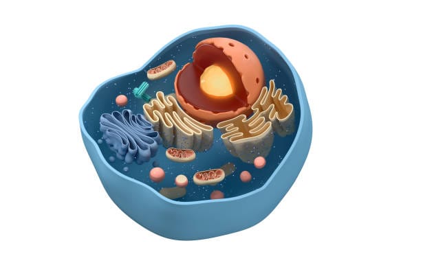
Inside each cell lies a complex network of molecular machinery. It’s not random or chaotic; it’s a system as deliberate as clockwork. Eukaryotic cells—found in plants, animals, fungi, and protists—are especially remarkable for the internal compartments they contain. These compartments are the organelles. They are separated from the rest of the cell by membranes, allowing them to maintain distinct environments and perform specialized functions with incredible efficiency.
Prokaryotic cells, such as bacteria, don’t have membrane-bound organelles. But even they exhibit a surprising level of organization with structures like ribosomes and nucleoid regions. Still, it’s in eukaryotic cells where the full orchestra of organelles comes to life, and where the interplay between structure and function becomes a masterpiece of biological design.
The Nucleus: The Library of Life
At the heart of the eukaryotic cell lies the nucleus, often considered the control center or command headquarters. Encased in a double membrane with carefully regulated pores, the nucleus safeguards the cell’s most precious possession: its DNA. This genetic material is like a vast instruction manual, a blueprint for every function the cell performs and every structure it builds.
Inside the nucleus, DNA is organized into chromatin—threads of genetic code that wind around proteins like spools. During cell division, chromatin condenses into chromosomes, ensuring accurate transmission of genetic information. Within this dense matrix sits the nucleolus, a smaller but vital structure responsible for assembling ribosomal subunits—the first step in protein construction.
The nucleus doesn’t operate in isolation. Messenger RNA (mRNA) molecules are transcribed from DNA and then transported out of the nucleus through nuclear pores. These messages are carried like sealed letters to the ribosomes, where they are translated into proteins. In this way, the nucleus is not merely a static storage space, but an active, responsive command center sending out precise instructions moment by moment.
Mitochondria: The Power Plants of the Cell
If the nucleus is the cell’s command center, then mitochondria are its power plants. These bean-shaped organelles are often described as the “powerhouses of the cell” for good reason: they are the sites of cellular respiration, the process by which energy is extracted from nutrients and converted into adenosine triphosphate (ATP), the molecule that powers nearly every activity in the cell.
Mitochondria have their own DNA—a remnant of their ancient origin as free-living bacteria that entered into a symbiotic relationship with early eukaryotic cells. This theory, known as endosymbiosis, explains many features of mitochondria: their double membrane, their independent replication, and their bacterial-like ribosomes.
Inside mitochondria, glucose and oxygen are chemically processed in a series of reactions known as the citric acid cycle and oxidative phosphorylation. The result is the generation of massive quantities of ATP, which is then distributed throughout the cell to power movement, synthesis, and signaling.
Mitochondria are also involved in other essential functions: they help regulate cell death (apoptosis), produce certain hormones, and even play roles in the immune response. When they malfunction, the consequences can be severe—ranging from muscle weakness to neurodegenerative diseases. In essence, the health of mitochondria is the health of the organism.
The Endoplasmic Reticulum: Cellular Highway and Factory
The endoplasmic reticulum (ER) is a sprawling network of membranes that extends from the nuclear envelope throughout the cytoplasm. It comes in two varieties—rough and smooth—and each plays a different but complementary role.
The rough ER is studded with ribosomes on its surface, giving it a bumpy appearance. These ribosomes are the sites of protein synthesis. As nascent proteins emerge from the ribosomes, they are folded and modified within the ER’s interior. Some will be shipped to the cell membrane, others to different organelles, and still others secreted out of the cell entirely.
The smooth ER, lacking ribosomes, serves other vital functions. It synthesizes lipids, including phospholipids and cholesterol, which are essential for building membranes. It also plays a role in detoxifying harmful substances, especially in liver cells, and in storing calcium ions, critical for muscle contractions and cellular signaling.
Together, the rough and smooth ERs serve as the cell’s factory floor and distribution network, managing both the production and initial transport of critical molecules.
The Golgi Apparatus: Master of Modification and Distribution
Once proteins and lipids are synthesized in the ER, they are packaged into vesicles and transported to the Golgi apparatus—a series of flattened, stacked membranes that resemble a stack of pancakes. But the Golgi is no breakfast buffet; it is a biochemical processing plant.
Here, proteins are further modified—sugars may be added in a process called glycosylation, or phosphate groups may be attached. These modifications are not decorative; they are functional, determining where the protein will go and how it will behave.
After processing, the Golgi packages these molecules into new vesicles, labeling them for delivery. Some are sent to the plasma membrane, others to lysosomes or secreted outside the cell. In effect, the Golgi is the cell’s post office—sorting, packaging, and delivering the products necessary for life.
Lysosomes: The Cell’s Recycling Centers
Life at the cellular level is not only about building—it’s also about breaking down and rebuilding. This is the task of lysosomes, membrane-bound sacs filled with powerful enzymes capable of digesting nearly every type of biological molecule.
Lysosomes break down old or damaged organelles, cellular debris, and even engulfed pathogens like bacteria. This process is known as autophagy, meaning “self-eating.” It’s a form of cellular housekeeping that prevents the accumulation of waste and allows the recycling of valuable materials.
When lysosomes malfunction, the consequences are dire. Inherited disorders like Tay-Sachs disease are the result of missing or defective lysosomal enzymes, leading to the buildup of toxic substances and often resulting in severe neurological damage.
But under normal conditions, lysosomes are not destructive. They are essential. They ensure that the cell maintains balance, efficiency, and cleanliness—a lesson perhaps worth noting in the human world as well.
Peroxisomes: The Cellular Detox Units
Peroxisomes are often overlooked in basic biology texts, but they play a critical role in metabolism and detoxification. These small, membrane-bound organelles contain enzymes that catalyze oxidative reactions, producing hydrogen peroxide as a by-product—hence the name.
Hydrogen peroxide is highly reactive and potentially dangerous, but peroxisomes contain another enzyme, catalase, which quickly breaks it down into water and oxygen. This makes peroxisomes important players in protecting the cell from oxidative stress.
They also participate in the breakdown of very long-chain fatty acids, the synthesis of certain phospholipids, and even in converting toxic substances like alcohol into safer compounds. Like the liver in the body, peroxisomes are the guardians against molecular toxicity within the cell.
Ribosomes: The Protein Assembly Lines
Ribosomes are not membrane-bound, but they are critical organelles nonetheless. They are the molecular machines that build proteins by reading instructions carried by mRNA. Each ribosome consists of two subunits, which clamp onto the mRNA strand and translate its sequence into a chain of amino acids—the building blocks of proteins.
Ribosomes can be free-floating in the cytoplasm or attached to the rough ER. Free ribosomes typically synthesize proteins that remain in the cell, while those on the rough ER make proteins destined for export or for use in membranes.
The process is elegant and astonishingly fast. A single ribosome can assemble hundreds of amino acids into a functional protein in just a few minutes. Without ribosomes, the genetic code would be meaningless—silent blueprints with no builder to bring them to life.
The Cytoskeleton: Structure, Transport, and Movement
Cells are not static blobs. They have shape, strength, and even mobility, all thanks to the cytoskeleton—a dynamic network of protein fibers that crisscross the cytoplasm.
The cytoskeleton is composed of three main types of fibers: microfilaments, intermediate filaments, and microtubules. Microfilaments help with cell shape and movement. Intermediate filaments provide structural stability. Microtubules serve as highways for transporting organelles and vesicles and are also essential in cell division, where they form the mitotic spindle.
Motor proteins “walk” along these fibers, carrying cargo like vesicles or mitochondria to specific locations. This intracellular traffic system is vital for efficiency and organization. Disruption of cytoskeletal elements can lead to disease and dysfunction, including cancer metastasis and neurodegenerative disorders.
The Plasma Membrane: Border Control and Communication
Every cell is surrounded by a plasma membrane—a flexible but sturdy barrier made of phospholipid bilayers and embedded proteins. This membrane regulates what enters and leaves the cell, maintaining an internal environment distinct from the chaotic outside world.
The plasma membrane is not a static wall; it’s an active participant in cellular life. Proteins embedded in the membrane act as sensors, receptors, and gateways. They receive signals from hormones, neurotransmitters, and nutrients and transmit those messages inside, triggering cascades of biochemical activity.
Membrane proteins also play a role in immune recognition, allowing the body to distinguish friend from foe. This becomes especially important in organ transplants and autoimmune diseases, where the recognition system may go awry.
Chloroplasts: The Solar Panels of Plant Cells
In the green world of plants, chloroplasts are the energy engines. Like mitochondria, chloroplasts evolved from free-living organisms that entered into symbiosis with a host cell. They have their own DNA, double membranes, and ribosomes.
Chloroplasts are the site of photosynthesis, where sunlight, carbon dioxide, and water are transformed into glucose and oxygen. This process not only powers the plant itself but also fuels nearly all life on Earth. The oxygen we breathe is a gift from chloroplasts. The food we eat, directly or indirectly, originates with them.
Inside chloroplasts are structures called thylakoids, which contain chlorophyll—the pigment that captures sunlight. Photosynthesis occurs here in stages, involving light reactions and the Calvin cycle, a complex dance of energy transformation.
Vacuoles: Storage and Support
Vacuoles are membrane-bound sacs with a variety of functions depending on the cell type. In plant cells, the central vacuole is especially prominent, taking up most of the cell’s volume. It stores water, nutrients, and waste products and maintains internal pressure (turgor), which keeps the plant rigid.
In animal cells, vacuoles are smaller but still important for storing substances and isolating harmful materials. In single-celled organisms like paramecia, contractile vacuoles help expel excess water, maintaining internal balance.
Though often considered “empty,” vacuoles are anything but—they are storage units, defense centers, and hydraulic support structures all in one.
A Symphony of Life
The story of organelles is a story of interdependence. No organelle works alone. Like members of an orchestra, they harmonize their efforts to produce the music of life. Disruption in one part of the system can throw the whole out of tune, resulting in disease or death.
This is not merely a collection of facts; it is a window into the intricate choreography of biology. Every heartbeat, every thought, every breath you take is powered by these microscopic structures working in tandem with a precision that defies imagination.
Understanding organelles is not just for scientists. It is for anyone who marvels at the complexity of life, who wants to know how the spark of existence is maintained from moment to moment in every living cell.