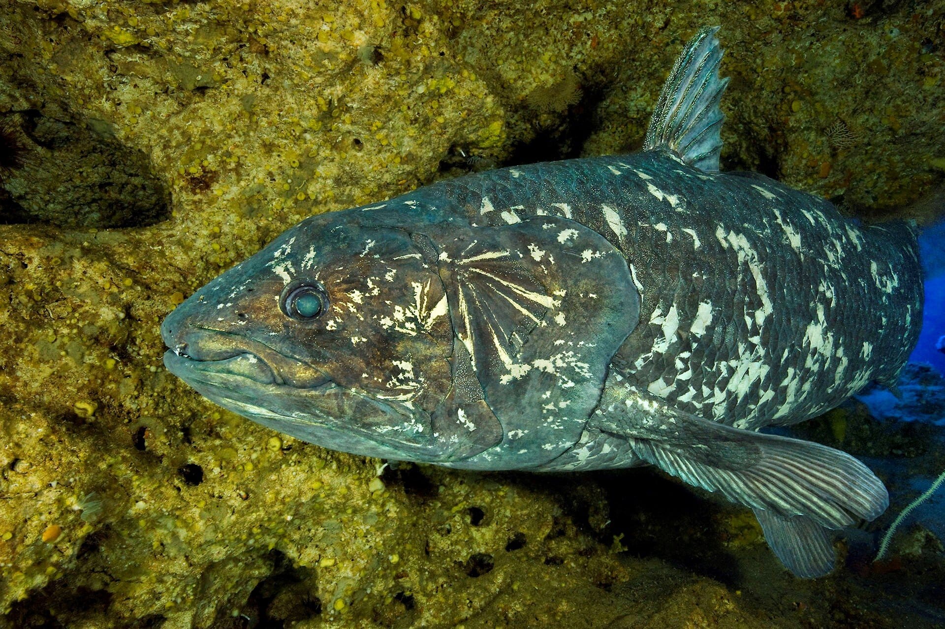
Beneath the crushing darkness of the ocean’s depths, in cold, ancient caves 300 meters below the surface, swims a fish that shouldn’t exist. With fleshy lobed fins, a bizarrely hinged skull, and a body plan seemingly untouched by time, the coelacanth has often been called a “living fossil.” But as it turns out, this label conceals more than it reveals. According to a groundbreaking new study published in Science Advances, this elusive fish is not merely a relic of the past—it is rewriting the very history of vertebrate evolution.
Led by Dr. Aléssio Datovo of the University of São Paulo (USP) and the late G. David Johnson of the Smithsonian Institution, the study offers a dramatic re-evaluation of one of the most intensively studied animals in the fossil record. By dissecting rare museum specimens of the African coelacanth (Latimeria chalumnae), the researchers have discovered that decades of scientific assumptions about its cranial muscles were wrong. And those mistakes may have clouded our understanding of how key anatomical features like jaws, breathing muscles, and feeding mechanisms evolved in the early branches of our own evolutionary tree.
As it turns out, the coelacanth has a few more secrets left to tell.
An Ancient Creature in a Modern Lab
The coelacanth is no ordinary fish. Once believed extinct for 65 million years, it was famously rediscovered alive off the coast of South Africa in 1938, shocking the scientific world. With origins stretching back over 400 million years, it represents a surviving lineage of lobe-finned fishes—the very group from which all land vertebrates (tetrapods) are descended. This makes the coelacanth more closely related to humans, birds, reptiles, and amphibians than it is to most other fish alive today.
Yet, despite its evolutionary importance, the coelacanth has remained frustratingly mysterious. They are rare, deep-sea dwellers, living in underwater caves, away from human eyes and access. Only a handful of specimens have ever been studied anatomically in detail, and many of the original anatomical descriptions relied heavily on assumptions and indirect observation.
Now, after years of persistence and patience, the researchers gained access to two extremely rare museum specimens from the Field Museum in Chicago and the Virginia Institute of Marine Science. With immense care, Datovo performed a full dissection—his life’s work condensed into six months of meticulous, microscopic effort.
“This was not just about cutting open a fish,” Datovo explains. “It was about peeling back the layers of 400 million years of evolution and correcting errors that have persisted in the literature for generations.”
Ligaments or Muscles? A Simple Mistake with Major Implications
At the heart of the study lies a crucial anatomical misunderstanding—one that has echoed through textbooks and scientific papers for decades.
In previous studies, scientists believed that coelacanths possessed a particular group of muscles inside their head—specifically, those responsible for expanding the buccopharyngeal cavity, which stretches from the mouth to the pharynx. These muscles are essential for suction feeding, a technique that allows fish to rapidly draw in prey by creating a vacuum inside their mouths. Ray-finned fish—such as goldfish, trout, and bass—are experts at this, and their suction abilities have contributed to their extraordinary evolutionary success. Today, ray-finned fish make up about half of all living vertebrates.
It was long assumed that these same muscles were also present in coelacanths, meaning they might have evolved in the common ancestor of all bony vertebrates. But the new study tells a very different story.
“Eleven structures that had been identified as muscles were actually ligaments—connective tissues that can’t contract,” says Datovo. “That has a drastic consequence for how this animal breathes and eats. Muscles create movement. Ligaments only transmit it.”
This finding changes everything. If coelacanths don’t have these suction-feeding muscles, then they couldn’t use suction to catch prey. Instead, like sharks and early jawed fishes, they rely on biting—a slower but mechanically powerful method of feeding.
More importantly, it pushes the origin of suction feeding 30 million years later than previously thought, suggesting it only appeared in the ancestor of modern ray-finned fish, not all bony vertebrates.
In other words, we’ve been imagining the wrong evolutionary sequence for decades.
A More Intimate Kinship with Sharks—and With Ourselves
Beyond debunking old anatomical errors, the study also sheds new light on the coelacanth’s evolutionary affinities. According to the researchers, coelacanths share more muscle structures with cartilaginous fishes—sharks, rays, and chimaeras—and tetrapods like amphibians and mammals, than with ray-finned fish.
That places them in a fascinating position on the vertebrate family tree. While they split from the ray-finned fish lineage some 420 million years ago, they represent a conserved model of what the early vertebrate head might have looked like before suction feeding and high-speed jaw mechanics took over the seas.
These findings also ripple forward through evolutionary time, altering how we interpret skulls and muscles in extinct species—including the first animals to crawl onto land.
Using three-dimensional microtomography scans of both living and fossilized fish skulls, Datovo and his colleagues reconstructed how the newly confirmed coelacanth muscles might have been arranged in ancient jawed vertebrates. This kind of comparative analysis opens the door to future work examining how structures like gill arches, cranial nerves, and respiratory muscles evolved into the complex systems we see in tetrapods today—including humans.
“This isn’t just about a fish,” says Datovo. “This is about understanding the evolutionary transformations that eventually gave rise to the human head.”
The Man Behind the Muscles
The study also carries a quiet emotional weight. G. David Johnson, a legendary ichthyologist and co-author of the paper, passed away in late 2024 while the manuscript was under peer review. His decades-long career produced some of the most detailed and influential studies on fish anatomy ever published.
“This paper is as much his legacy as it is a scientific achievement,” says Datovo. “He was probably the greatest fish anatomist of his time. Without his dedication, we wouldn’t have had access to these priceless specimens. His contribution lives on in every muscle we examined.”
Contrary to common belief, dissecting museum specimens does not destroy them if done correctly. In this case, every muscle, bone, and nerve was preserved separately and cataloged, allowing future scientists to study them without ever harming another coelacanth. In this way, Johnson’s final project becomes an enduring resource for decades of future research.
Rethinking Evolution Through a Ghost from the Deep
More than 85 years after its rediscovery, the coelacanth continues to confound, humble, and inspire scientists.
Living fossils are not static snapshots of the past. As this study proves, they are dynamic, complex, and evolving organisms—offering insights not because they haven’t changed, but because they have retained ancestral features that have otherwise vanished from modern lineages.
In peeling back the layers of the coelacanth’s skull, Datovo and Johnson have reminded us that even the most well-known icons of evolutionary biology still hold mysteries waiting to be uncovered. And that our understanding of life’s grand history is only as solid as the evidence we’re willing to examine—muscle by muscle, ligament by ligament.
In the coelacanth, we glimpse both the deep past and the enduring power of curiosity. Not just a relic, but a teacher. Not just a fossil, but a living witness to our own evolutionary origins. And, if we’re willing to listen closely, still whispering truths from the ancient sea.
Reference: Aléssio Datovo et al, Coelacanths illuminate deep-time evolution of cranial musculature in jawed vertebrates, Science Advances (2025). DOI: 10.1126/sciadv.adt1576