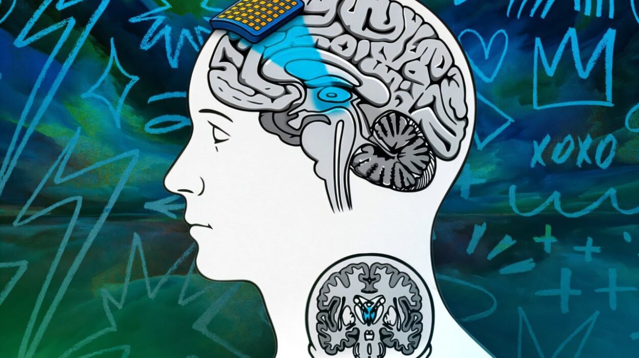
For decades, scientists have tried to peer into the intricate wiring of the brain, hoping to understand how its countless cells connect, communicate, and create the symphony of thought. Yet, even with the most advanced tools, much of this inner universe has remained hidden behind the limits of technology. Now, an international team led by the Francis Crick Institute, working with the Paul Scherrer Institute, has taken a bold step forward. Their new imaging protocol, published in Nature Methods, opens a window into mouse brain circuitry with detail so crisp it feels almost unreal—yet it is made possible by a surprising tool borrowed from the aerospace industry.
When Slices No Longer Suffice
For years, volume electron microscopy has been the gold standard for exploring the brain’s “circuitry.” The technique is powerful, but it comes at a steep price: biological tissue must be cut into extraordinarily thin slices, tens of thousands of them for every millimeter. Each slice is imaged, and the entire set is stitched back together like a 3D jigsaw puzzle. It works beautifully for small creatures like fruit flies—so well, in fact, that scientists have already built full connectomes of fruit fly larvae and adult flies. But when it comes to mammalian brains, the challenge balloons out of proportion. There is simply too much material to slice with reliability.
Andreas Schaefer, Principal Group Leader at the Crick, captures the frustration and excitement of the field in one breath. “Volume EM has been revolutionary for seeing things inside the cell in 3D, but it comes with limitations for mapping neuron connections inside mammalian brains, which are too large to be reliably sliced into tiny sections,” he said. “We’re excited that our protocol and use of powerful radiation-resistant material allowed brain tissue to be imaged at extraordinary resolution. Refining this technique further could bring us one small step closer to a future goal in the field: mapping the mouse brain connectome, which is tens of thousands of times bigger than the fruit fly connectome.”
The question, then, was clear: could researchers find a way to bypass the slicing altogether?
Borrowing Strength from Spacecraft
X-rays offered a tantalizing alternative. Unlike electrons, X-rays can penetrate deeply into matter. The team wondered whether the right preparation could allow X-rays to capture delicate neuronal details without damaging the tissue. But raw biological samples could not withstand the intense radiation needed to illuminate the microscopic structures.
So the researchers did something unexpected. They turned to materials originally designed to withstand the punishing conditions inside nuclear reactors and spaceships. Building on standard sample preparation protocols, they embedded stained brain tissue in a special radiation-resistant resin—a substance tough enough to protect machines built for the extremes of Earth and beyond.
This resin changed everything. It allowed the samples to endure at least twenty times more radiation than usual, tolerating “billions more X-rays than would be fatal for a human.” What would have previously destroyed the tissue now left it perfectly intact, ready for imaging at a scale never before achieved with X-rays.
Riding the Synchrotron’s Light
To take advantage of this newfound durability, the team brought their samples to a synchrotron, a massive particle accelerator where electrons race at near-light speed. Guided by magnetic and electric fields, those electrons emit intense, coherent X-rays—light powerful enough to uncover structures hidden deep within solid matter.
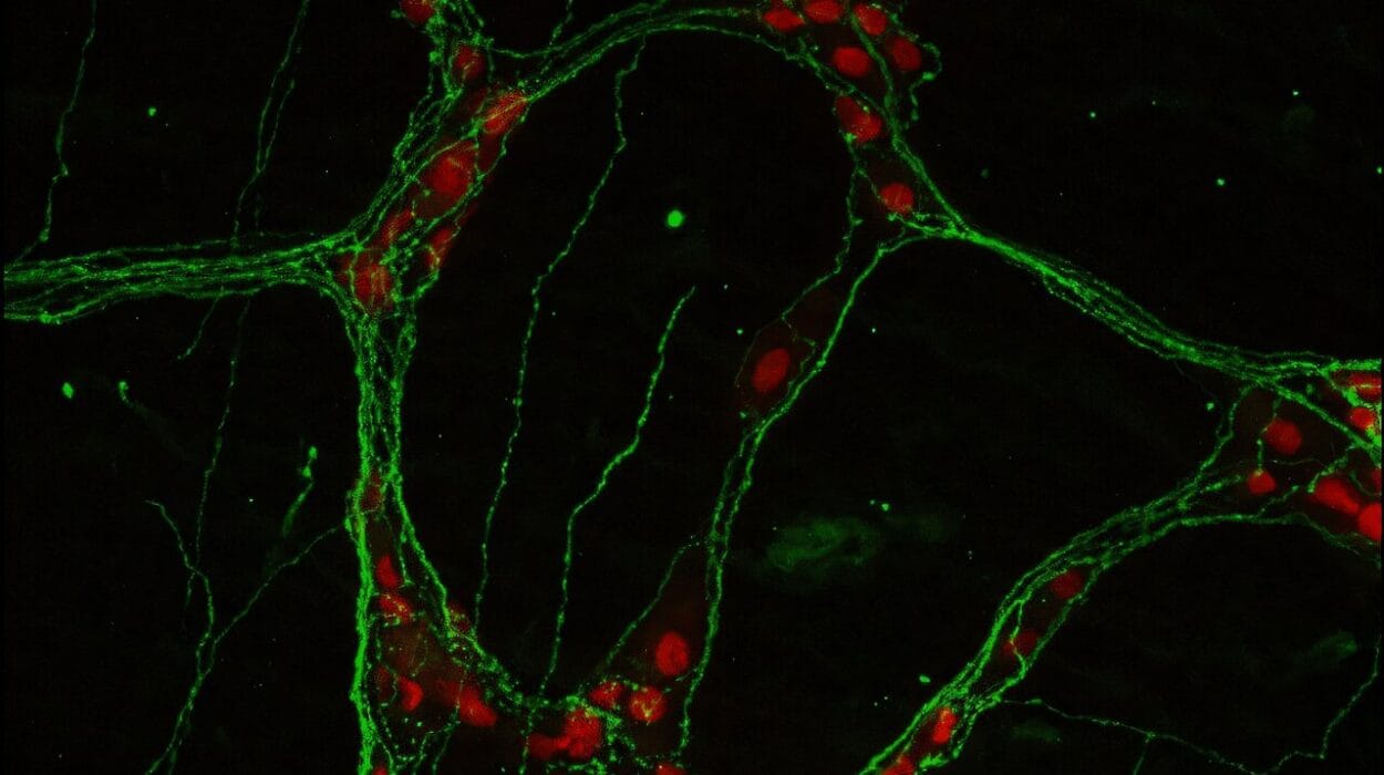
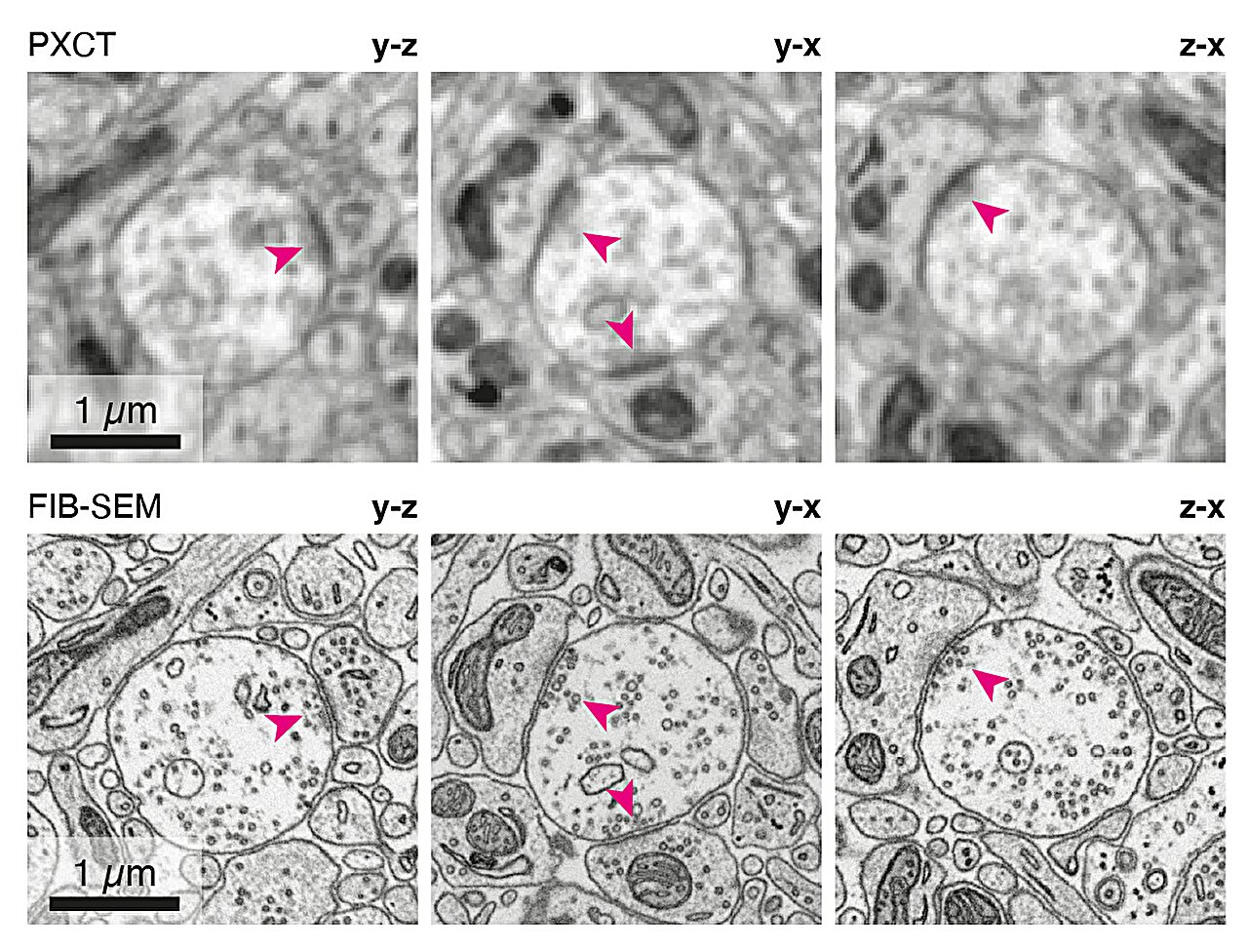
The imaging technique they used, X-ray ptychography, allowed them to reconstruct the tissue’s internal architecture with extraordinary precision. When the images appeared, the researchers could hardly believe what they were seeing. Synapses, dendrites, axons—the essential components of neural communication—came into view at a resolution of 38 nanometers. For the first time, these microscopic features could be observed in 3D without slicing the tissue at all.
A Future Built on Clearer Views
The breakthrough was not just about proving that X-rays could work. It was about expanding what is possible for brain imaging as a whole. Carles Bosch Piñol, Principal Laboratory Research Scientist at the Crick, expressed the team’s vision. “Now we’ve shown that X-ray imaging is suitable for mapping the fine detail of delicate biological tissue samples in 3D, we’re continuing to make the methods better and better,” he said. “We want to improve the field of view, addressing larger samples, and the resolution, obtaining finer details. Combining X-ray imaging with other methods opens up new possibilities to study the function of biological tissues such as the brain.”
The project was jointly led by Ana Diaz and Adrian Wanner at the Paul Scherrer Institut in Switzerland, with key contributions from the European Synchrotron Radiation Facility and the Electron Microscopy team at the Crick. Together, these collaborators have created a foundation that promises to reshape the study of neural architecture.
Why This Breakthrough Matters
The brain is often compared to a universe contained within a skull—complex, vast, and full of mysteries. Mapping its connectome, especially in mammals, is one of the most ambitious scientific quests of our time. Every step toward clearer imaging brings us closer to understanding how thoughts form, how memories are stored, how diseases disrupt neural communication, and how this remarkable organ orchestrates life itself.
This new imaging protocol represents more than a technical improvement. It is a conceptual shift, a demonstration that tools forged for the harshest corners of human engineering—from reactors to spacecraft—can reveal the delicate inner world of the brain. By allowing X-rays to reach the necessary depth and detail without destroying the tissue, the technique opens a path to study the mammalian connectome with unprecedented clarity.
As researchers refine and expand this method, the dream of a full mouse brain map moves from the realm of the impossible toward the achievable. And with every neuron illuminated and every connection traced, we take another step toward unraveling the profound mysteries of the mind.
More information: Nondestructive X-ray tomography of brain tissue ultrastructure, Nature Methods (2025). DOI: 10.1038/s41592-025-02891-0