Scientists have uncovered an unexpected transport route neurons use to move critical receptors across their unusually long axons—a process essential for keeping synaptic communication working. Using advanced imaging, researchers found that the TrkA receptor often takes an indirect pathway called transcytosis, traveling to the cell surface before being pulled back inside and shipped down the axon. Disrupting this route in mice reduced presynaptic structures and weakened synaptic transmission, revealing how vital this hidden system may be for brain function.
All cells depend on proteins to survive and function. But neurons face a challenge most other cells never encounter: distance.
A neuron’s axon—the thin, thread-like structure responsible for carrying electrical impulses—can extend for meters. That means proteins made in the cell body, or soma, may need to travel enormous lengths before reaching the far end of the cell. For neurons to maintain normal communication, they must constantly deliver the right materials to the right place at the right time.
A new study published in Science Signaling reveals that neurons solve part of this problem using an unconventional delivery strategy—one that researchers are only now beginning to fully visualize.
Two pathways, but one remained mysterious
Scientists have long known that neurons can ship proteins down axons through a more “standard” intracellular route.
In this pathway, newly made proteins are packaged in the trans-Golgi network and sent outward through the secretory pathway, moving directly toward their destination.
But neurons also use a second method called transcytosis, and until now, much of this process remained unclear. Researchers had limited understanding of how fast it works, what structures carry proteins, and how the neuron decides when to use it.
That knowledge gap mattered, because neurons rely on precise protein delivery to keep synapses functioning—especially at the presynaptic terminals where neurotransmitters are released.
Tracking a critical receptor across the neuron
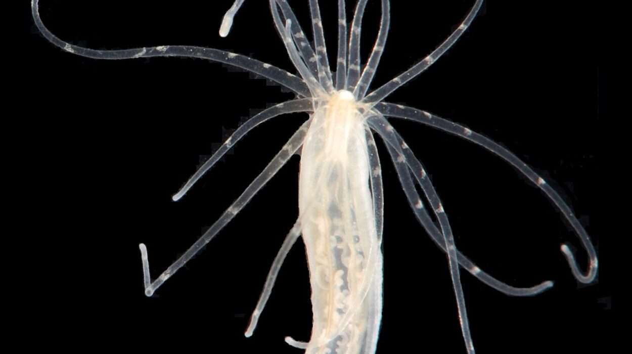
To investigate this hidden route, researchers focused on the TrkA (tropomyosin-related kinase A) receptor, part of the Trk receptor family. TrkA is especially important because it influences how neurons grow, survive, and communicate.
It does this by responding to a signal called nerve growth factor (NGF). When NGF binds to TrkA at the far end of the axon, the receptor carries that signal back toward the cell body, helping deliver essential information about what the neuron needs.
Because TrkA naturally travels long distances, it became an ideal candidate for studying how neurons manage transport across extended axons.
A microfluidic setup that revealed the route
To watch TrkA movement in real time, the researchers grew mouse nerve cells in specially designed microfluidic chambers. These chambers allowed the team to separate the neuron’s cell body region from its axon region, creating distinct compartments.
This design gave the researchers precise experimental control. They could tag receptors located on the soma side with fluorescent markers while independently applying NGF only to the axon side.
That setup made it possible to track how receptors responded when the axon “requested” more supplies.
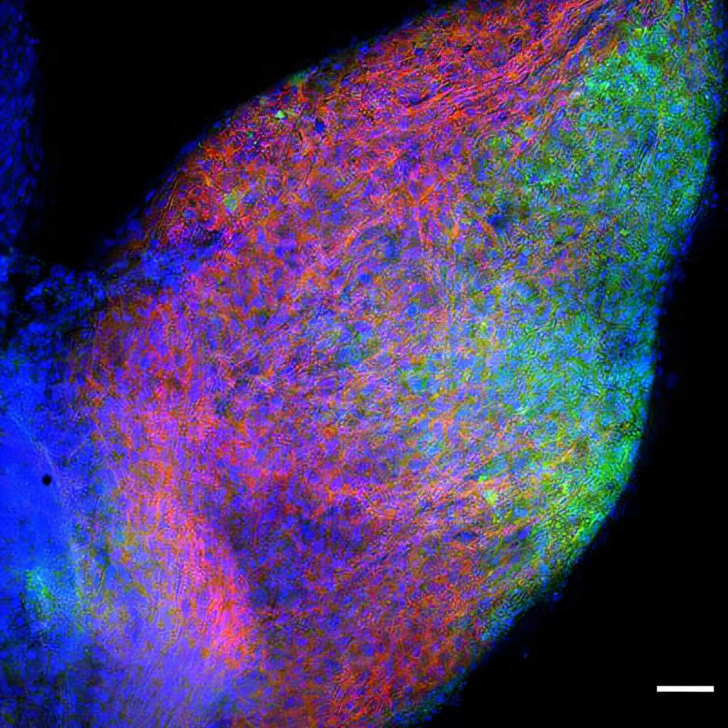
Using a high-resolution electron microscope, the team followed the labeled receptors as they moved across the neuron.
The discovery: TrkA doesn’t always go straight to the axon
The imaging revealed something unexpected.
The TrkA receptor does not always take the direct route from the cell body to the axon. Instead, it often takes a transcytosis detour. In this pathway, TrkA first travels to the surface of the soma, then gets pulled back inside the neuron, packaged into vesicles, and transported down the axon.
This indirect system may seem inefficient at first glance, but the researchers found it appears to be regulated by signals coming from the axon itself.
In other words, the axon can influence what the soma sends and when.
NGF triggers a feedback loop for protein delivery
The researchers found that this transcytosis route is activated by NGF arriving from the far end of the neuron.
That means the axon is not passively receiving materials—it is actively communicating its needs. NGF triggers a positive feedback loop, signaling the cell body that additional supplies of critical receptors are needed.
The result is a dynamic delivery mechanism where the neuron adjusts its internal shipping based on demand at distant synaptic sites.
This finding helps explain how neurons maintain function despite extreme internal distances.
The cellular “vehicles” that carry receptors down the axon
The study also clarified what physical structures carry transcytosed receptors.
Microscopy revealed that receptors travel inside small membrane-bound compartments, including endosomes and multivesicular bodies. These compartments act as transport containers, allowing receptors to move safely through the axon’s internal environment.
The researchers also identified the motor protein responsible for moving these cargo units: KIF1A.
KIF1A carries the receptor-filled compartments along the axon, driving long-range transport toward presynaptic regions.
Delivery targets: presynaptic varicosities
One of the most important findings was where these receptors end up.
The team observed that transcytosed TrkA receptors are transported specifically to presynaptic varicosities—small swellings along axons where neurotransmitters are released.
These varicosities play a central role in synaptic signaling. By delivering TrkA receptors directly to these locations, the neuron may ensure that synaptic communication remains functional and responsive.
This suggests transcytosis is not a random detour, but a targeted system designed to support specific synaptic structures.
What happens when transcytosis is disrupted
To test whether this pathway is truly necessary, the researchers genetically modified a mouse model to interfere with the transcytosis route.
The effects were clear.
When transcytosis was disrupted, presynaptic sites became fewer and smaller, and synaptic transmission weakened. This indicates that the transcytosis pathway is not merely a backup system—it plays a direct role in maintaining the physical and functional integrity of synaptic release sites.
The findings provide strong evidence that long-distance protein delivery is tightly linked to synaptic strength.
Why this matters
Understanding how neurons transport proteins is not just a question of basic biology—it is central to understanding how the nervous system stays functional over a lifetime.
This study reveals that neurons rely on an atypical but essential delivery system—transcytosis—to ensure critical receptors like TrkA reach presynaptic regions where communication depends on them. The discovery that this pathway involves endosomes, multivesicular bodies, and the motor protein KIF1A, and that disrupting it weakens synaptic transmission, provides a clearer mechanistic view of how neurons maintain connectivity.
Because proper protein transport is fundamental to nerve repair and neuronal stability, these insights could help reshape how researchers think about treating nerve damage from injury or disease, as well as disorders linked to disrupted neuronal connectivity and neurodegeneration.
Study details
Guillermo Moya-Alvarado et al, Transcytosis-mediated anterograde transport of the receptor TrkA mediates the formation of presynaptic sites in sympathetic neurons, Science Signaling (2026). DOI: 10.1126/scisignal.aea7078