Depression is associated with measurable thinning in several regions of the brain’s cortex, according to one of the largest MRI analyses conducted to date. Examining brain scans from more than 12,000 people, researchers found that these structural differences vary with age, disease stage, and antidepressant use, offering a clearer picture of the disorder’s biological signatures while highlighting the need for further validation before clinical use.
Depression affects millions of people worldwide, yet many of its biological foundations remain difficult to pin down. Scientists have long suspected that major depressive disorder leaves measurable traces in the brain, but identifying consistent structural patterns has proven challenging.
Now, one of the largest brain imaging analyses of depression has revealed that people with major depressive disorder (MDD) consistently show reduced cortical thickness across several brain regions. The findings, based on MRI scans from more than 12,000 individuals, suggest that these changes are influenced by a person’s age, whether they are experiencing an active depressive episode, and, to a lesser extent, whether they are taking antidepressant medications.
The research, published in Nature Mental Health, provides what the authors describe as a high-resolution map of brain structure differences linked to depression.
Thousands of Brain Scans Combined Into a Single Global Analysis
To better understand the brain’s structural characteristics in depression, researchers from the Chinese Academy of Sciences and Vrije Universiteit Amsterdam analyzed MRI scans from 5,736 people diagnosed with MDD and 6,538 individuals with no known psychiatric disorders.
Rather than relying on data from a single hospital or research center, the team combined brain scans collected by 64 independent research groups worldwide. The datasets came from two international collaborations: the Enhancing NeuroImaging Genetics through Meta-Analysis (ENIGMA) MDD consortium and the Depression Imaging Research Consortium (DIRECT).
The researchers processed every MRI scan using the same standardized methods, allowing them to compare brain structures across thousands of participants with greater consistency.
Instead of examining the brain only at broad regional levels, they performed a vertex-wise (point-by-point) meta-analysis, enabling a detailed assessment of the cerebral cortex.
Depression Was Linked to Thinner Cortical Regions
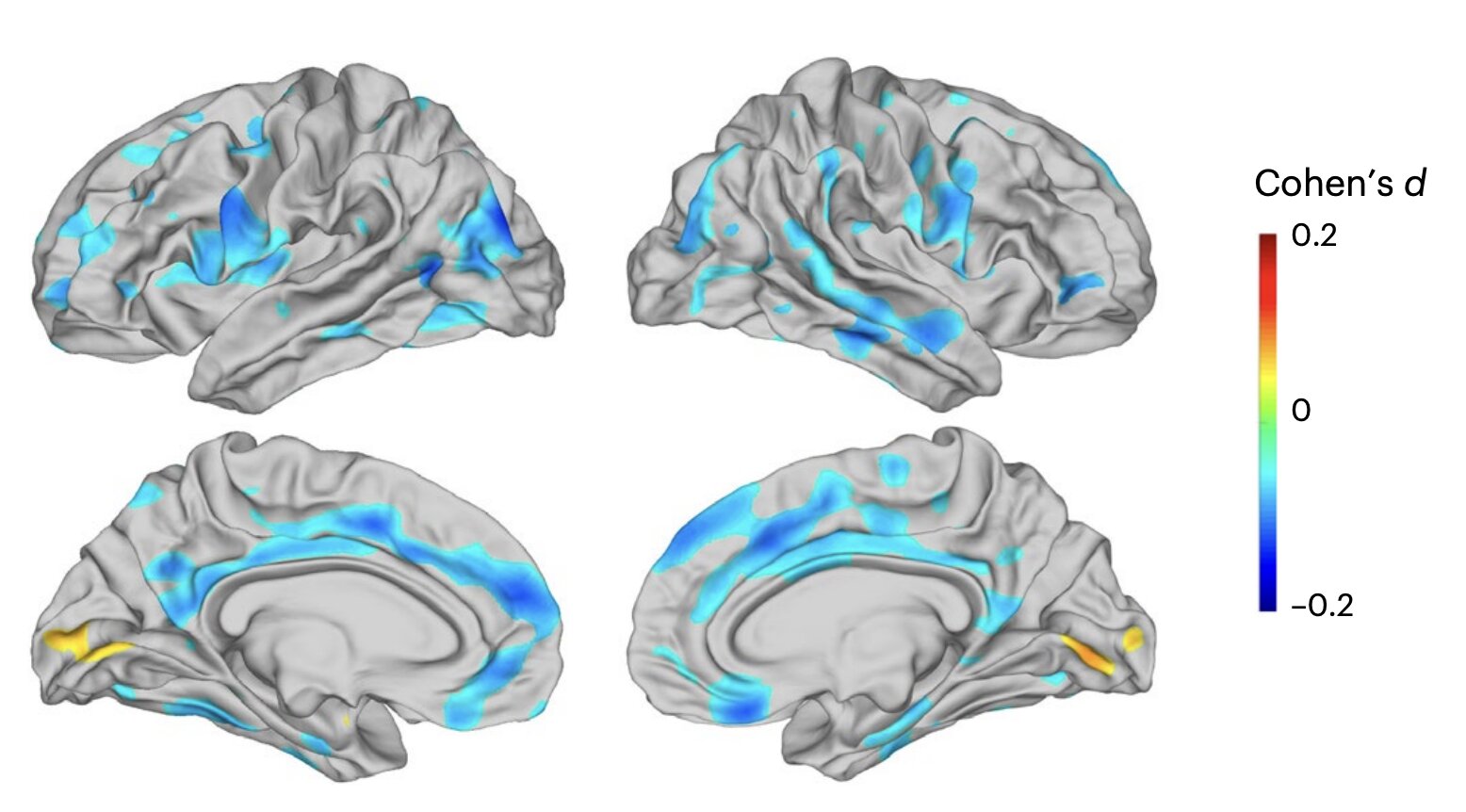
The analysis uncovered a consistent pattern: people with major depressive disorder had lower cortical thickness in multiple parts of the brain.
These differences appeared in regions including the inferior parietal, lateral occipital, superior parietal, medial and lateral orbitofrontal, anterior and posterior cingulate, and precentral gyri.
While cortical thickness differed between the two groups, the researchers found no significant differences in cortical surface area. This suggests that depression is associated with changes in the thickness of the cortex rather than its overall size.
The results provide a more refined picture of how depression may be reflected in brain anatomy, although the study does not establish whether these structural differences cause depression or result from it.
Age and Disease Stage Made a Difference
The brain differences were not equally apparent across every participant.
The researchers found that cortical thinning was more pronounced in adults who were experiencing an acute depressive episode. In contrast, adolescents with depression did not show significant structural differences compared with adolescents who had no known mental health disorders.
These findings suggest that the brain-related signatures of depression may evolve over time or become more detectable during specific stages of the illness.
The study highlights that depression is unlikely to have a single, uniform pattern of brain changes across all age groups.
Antidepressant Use Showed Only Modest Effects
The researchers also examined whether antidepressant medications were associated with differences in brain structure.
Patients taking medications such as selective serotonin reuptake inhibitors (SSRIs) and serotonin and norepinephrine reuptake inhibitors (SNRIs) showed slightly greater cortical thinning than patients who were not taking these drugs.
However, the researchers emphasized that these effects were subtle and modest in size. The findings do not indicate that antidepressants cause these structural differences, only that the observed brain patterns were somewhat more noticeable among medicated patients within this analysis.
Building Better Models of Depression
Although the findings are not ready for clinical application, they provide a more comprehensive framework for understanding depression’s structural characteristics.
According to the researchers, this globally derived, high-resolution brain map could support future studies investigating the biological mechanisms behind depression. It may also help scientists evaluate potential structural markers related to disease progression and responses to treatment.
The standardized approach across dozens of research groups strengthens confidence that the observed patterns are not limited to a single population or imaging center.
Why This Matters
Depression remains one of the world’s most common psychiatric disorders, yet its biological signatures have been difficult to define consistently. By analyzing MRI scans from more than 12,000 people, this study identifies reproducible patterns of cortical thinning that appear to distinguish adults with major depressive disorder from people without known psychiatric conditions.
The researchers stress that these findings are preliminary and should not be used to diagnose or assess patients. Additional studies will be needed to confirm whether these structural differences can reliably serve as biomarkers. If future research validates these results, they could contribute to improved methods for studying depression, monitoring its clinical course, and eventually developing more precise diagnostic or treatment assessment tools.