An injectable hydrogel loaded with extracellular vesicles from IL-4/C1q-activated astrocytes rebuilt blood vessels inside stroke-damaged brain cavities, attracted reparative immune cells, and restored motor function in mice to nearly healthy levels. Instead of focusing only on surviving brain tissue around a stroke, the approach targeted the long-neglected dead core of the injury, suggesting a potential strategy for repairing tissue after the window for emergency stroke treatment has already closed.
For decades, the dead center of an ischemic stroke has been viewed as one of the greatest obstacles in brain repair. Once brain tissue dies, it leaves behind a cavity that current medical treatments cannot rebuild. Emergency therapies such as clot-dissolving drugs and mechanical clot removal can reduce damage if given quickly, but many patients miss this narrow treatment window. Afterward, rehabilitation helps the brain adapt, but it cannot replace tissue that has already been lost.
The new research takes aim at that long-standing challenge. Rather than trying to rescue dying brain cells during the earliest hours after a stroke, the scientists developed a way to transform the empty cavity left behind into an environment capable of supporting regeneration.
Their strategy combines a specially engineered microporous annealed particle scaffold (MAPS) with tiny biological packages called extracellular vesicles (EVs) released by activated astrocytes, the abundant support cells of the brain. Together, the scaffold and the vesicles created a regenerative environment that recruited helpful immune cells, rebuilt blood vessels, encouraged nerve fiber growth, and ultimately restored movement in mice.
Turning an Empty Stroke Cavity Into a Healing Environment
The researchers designed an injectable granular hydrogel made from microscopic particles of hyaluronic acid, a naturally occurring molecule found in the body. When injected into the stroke cavity five days after stroke, the particles assembled into a porous scaffold.
Unlike conventional hydrogels that form a continuous gel, this scaffold contains interconnected spaces large enough for cells to move through immediately. Those pores were central to the design because they allowed immune cells and other repairing cells to enter the damaged region.
The team then attached extracellular vesicles directly onto the surfaces of these microscopic particles.
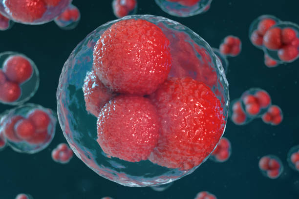
Extracellular vesicles are tiny membrane-bound particles that cells naturally release. They carry proteins, RNA molecules, and other biological signals capable of influencing distant cells.
Instead of floating freely and rapidly dispersing, the vesicles were chemically anchored to the scaffold. This allowed the biological signals to remain inside the damaged brain tissue long enough for infiltrating cells to encounter them repeatedly.
The researchers believed this prolonged local presentation might succeed where simply injecting vesicles had shown only limited benefit.
Two Types of Activated Astrocytes Produced Very Different Vesicles
Astrocytes can adopt different biological states after injury.
To investigate whether these different states influence healing, the researchers generated three populations of rat astrocytes in the laboratory.
One group remained untreated.
A second group was activated using IL-1α, TNF-α, and C1q, producing a state resembling inflammatory reactive astrocytes.
The third group received IL-4 together with C1q, creating what the researchers describe as a pro-repair astrocyte state.
RNA sequencing confirmed that the activated astrocytes developed distinct gene-expression profiles.
The scientists then isolated extracellular vesicles from each astrocyte population.
Importantly, all three groups produced vesicles with similar sizes—roughly 50 to 150 nanometers—and similar particle concentrations of about 1 × 10⁷ particles per milliliter. This meant the cytokine treatments did not substantially alter how many vesicles astrocytes released. Instead, they changed what those vesicles carried.
That difference in cargo proved to be crucial.
Injecting Vesicles Alone Was Not Enough
Before combining the vesicles with the scaffold, the researchers tested whether simply injecting IL-4/C1q-derived extracellular vesicles into the stroke cavity could repair the injury.
It did not.
Although the free vesicles produced modest improvements in nerve fiber preservation around the damaged region, they failed to regenerate the infarct core or substantially improve recovery.
This finding matched previous work suggesting that extracellular vesicles alone often disperse too quickly to remain effective.
The researchers therefore reasoned that keeping the vesicles physically anchored inside the damaged tissue might dramatically improve their therapeutic impact.
The Scaffold Restored Movement Nearly to Healthy Levels
To test their idea, the team used a photothrombotic stroke model in mice that produces lasting forelimb movement problems.
Five days after stroke, mice received one of several treatments.
Some received the scaffold alone.
Others received scaffolds carrying extracellular vesicles from untreated astrocytes.
Another group received scaffolds carrying vesicles from inflammatory astrocytes.
The final group received scaffolds carrying vesicles from IL-4/C1q-activated astrocytes.
Motor function was tracked for months using a grid-walking task that measures how often mice miss their footing.
Immediately after stroke, every treatment group showed similarly severe impairment.
But beginning about 16 days after stroke, only the mice receiving IL-4/C1q EV-loaded scaffolds showed a significant reduction in movement errors.
Their recovery continued over time.
By approximately eight weeks after stroke, their performance had improved until it was nearly indistinguishable from healthy animals.
None of the other treatments achieved similar recovery.
The Scaffold Became a Living Part of the Healing Tissue
The porous scaffold was never intended to remain unchanged.
Instead, it served as a temporary framework that cells gradually remodeled.
The researchers found that scaffolds carrying extracellular vesicles degraded more rapidly than scaffolds without vesicles.
As cells entered the porous spaces, they expanded those voids while breaking down the surrounding material.
This remodeling was not simply a mechanical consequence of attaching vesicles to the scaffold.
Instead, the results suggested that the vesicles actively attracted cells and strengthened interactions between those cells and the biomaterial, accelerating reconstruction of the damaged region.
New Blood Vessels Grew Deep Into the Stroke Core
One of the most striking findings involved rebuilding the brain’s circulation.
Normally, virtually no functioning blood vessels exist in the center of a mature stroke cavity.
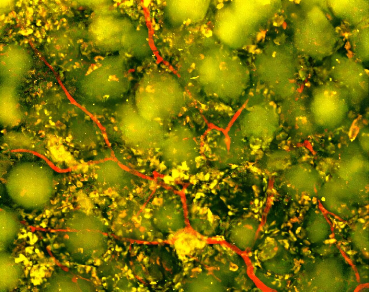
The researchers injected Tomato Lectin shortly before examining the brains, allowing them to visualize only vessels that were actively connected to the bloodstream.
Scaffolds carrying vesicles from IL-4/C1q-activated astrocytes generated robust networks of perfused blood vessels extending deep into the infarct.
Scaffolds carrying vesicles from inflammatory astrocytes also stimulated blood vessel growth, but the vessels formed after IL-4/C1q treatment occupied a larger area and developed longer branches, indicating stronger angiogenic activity.
Additional staining showed early recruitment of pericytes, cells that stabilize blood vessels.
Although these new vessels had not yet reached the maturity seen in normal brain tissue, their development represented an important step toward rebuilding functional circulation inside previously dead tissue.
The researchers linked this rapid vascular reconstruction to the dramatic improvements in motor recovery.
Nerve Fibers Also Returned, but Only One Treatment Restored Functional Circuit Remodeling
Growing nerve fibers alone does not necessarily restore brain function.
The researchers therefore examined both general axonal growth and specific motor pathways.
Both activated vesicle treatments increased the amount of NF200-positive nerve fibers within and around the infarct.
Interestingly, vesicles from inflammatory astrocytes actually produced more overall axonal coverage.
However, that increase did not translate into improved movement.
To understand why, the scientists repeated the experiments using mice whose motor and sensory neurons naturally fluoresce.
These animals revealed an important difference.
Only the IL-4/C1q treatment promoted robust regrowth of labeled motor and sensory nerve fibers in tissue surrounding the infarct.
The treated brains also regained nearly normal structural symmetry between the two hemispheres, indicating reduced tissue distortion after stroke.
The researchers emphasized that additional future studies using markers of neural activity and synaptic connections will be necessary to determine exactly how completely these new fibers restore functional communication.
The Therapy Recruited Helpful Immune Cells Instead of Triggering Harmful Inflammation
One of the central discoveries emerged from single-cell RNA sequencing, which profiled thousands of individual cells inside the injured brain.
Rather than suppressing the immune system, the therapy reshaped it.
Compared with scaffolds alone, extracellular vesicle-loaded scaffolds substantially increased infiltration of monocytes, macrophages, and neutrophils while reducing the dominance of resident inflammatory glial cells.
The IL-4/C1q treatment produced particularly large increases in reparative immune populations.
It also expanded endothelial cells, which form blood vessels, and cells belonging to the oligodendrocyte lineage, suggesting that multiple regenerative cell types accumulated together within the scaffold.
The therapy also shifted the balance of T-cell populations.
Overall T-cell numbers declined, but potentially damaging CD8 effector T cells became less common while regulatory T cells, Th17 cells, and γδ T cells became more abundant.
Computational analysis suggested that regulatory T cells were associated with vascular remodeling, anti-inflammatory signaling, and tissue repair, while γδ T cells were linked primarily to angiogenesis.
Together, these changes indicated that the scaffold was creating a fundamentally different immune environment inside the damaged brain.
Neutrophils Turned Out to Be Essential
Neutrophils are often viewed as destructive during the earliest stages after stroke because they contribute to inflammation.
But this study revealed a different role later in recovery.
The researchers found that IL-4/C1q EV-loaded scaffolds recruited the largest numbers of neutrophils.
Gene-expression analyses suggested these neutrophils possessed strong pro-angiogenic characteristics.
Predicted communication networks also identified multiple signaling pathways linking neutrophils and endothelial cells involved in blood vessel formation.
To determine whether neutrophils were actually responsible for repair, the researchers depleted them using antibodies.
The consequences were dramatic.
Removing neutrophils sharply reduced blood vessel growth and significantly decreased scaffold remodeling.
These experiments demonstrated that neutrophils were not merely present—they were required for much of the regenerative process observed after treatment.
Reparative Macrophages Added Another Layer of Healing
The study also examined different macrophage populations.
The researchers identified a subset expressing genes associated with tissue repair, including Spp1, Gpnmb, Lgals3, and Cd63.
This reparative macrophage population was most abundant in mice treated with IL-4/C1q EV-loaded scaffolds.
Additional experiments showed that these vesicles attracted macrophages more effectively than other vesicle types.
The scientists also found evidence suggesting that incoming monocyte-like cells may transform into reparative macrophages after entering the injured brain under the influence of the vesicles.
Communication analyses predicted multiple signaling pathways through which macrophages could promote new blood vessel formation.
Together with neutrophils, these macrophages appeared to create an immune environment favorable for rebuilding damaged tissue.
Small RNA Molecules May Explain Why IL-4 Made Such a Difference
To understand why IL-4/C1q-derived vesicles worked better than the others, the researchers analyzed the small RNA molecules carried inside them.
The vesicles possessed a distinct collection of microRNAs compared with vesicles from untreated or inflammatory astrocytes.
Among the most notable were miR-377-3p and miR-143-5p, whose predicted target genes were enriched in macrophages and neutrophils.
The authors propose that these molecules may help strengthen immune-cell adhesion and support recruitment of reparative leukocytes into the injured brain.
The analysis also showed enrichment of microRNAs associated with Wnt signaling, TGF-β signaling, axon guidance, Rap1 signaling, and cell-adhesion pathways, all of which are connected with tissue repair.
To determine whether C1q alone could generate the same effect, the researchers tested vesicles from astrocytes exposed only to C1q.
Those vesicles failed to stimulate comparable blood vessel growth, demonstrating that IL-4 activation was necessary to produce the regenerative vesicle cargo.
Why the Findings Matter—and What Comes Next
The researchers argue that their work shifts attention toward rebuilding the stroke infarct itself rather than focusing exclusively on surviving tissue around it.
Instead of relying on transplanted cells, the approach uses a cell-free biomaterial carrying naturally produced extracellular vesicles to organize the body’s own repair mechanisms.
By attracting reparative immune cells into a porous scaffold placed directly inside the stroke cavity, the treatment transformed what has long been considered an irreparable void into a regenerative niche.
The authors also acknowledge several important limitations.
Although neutrophil depletion established a key role for those cells, the precise contributions of different macrophage populations remain incompletely understood. The extracellular vesicles were isolated using a precipitation-based method that may include non-vesicle material, although every experimental group underwent identical processing. The researchers also note that they did not perform the full range of characterization recommended by the latest extracellular vesicle guidelines. Finally, while the temporary opening of the blood-brain barrier allows immune cells to enter the injured brain after stroke, the exact mechanisms governing how those cells are recruited and retained inside the scaffold still need to be clarified.
Future work will focus on refining the biological cargo carried by extracellular vesicles, exploring additional cell sources, understanding how immune responses change throughout stroke recovery, and testing the therapy in larger animal models while carefully evaluating long-term safety.
Although the treatment remains far from clinical use, the study demonstrates that rebuilding the dead center of a stroke may be more achievable than previously thought. By combining an engineered scaffold with carefully programmed biological signals, the researchers showed that the injured brain can recruit its own repair machinery to regenerate blood vessels, remodel tissue, and restore movement long after the initial stroke has passed.
More information
Shangjing Xin et al, IL-4/C1q activated astrocyte-derived extracellular vesicles promote stroke infarct recovery by recruiting peripheral leukocytes, Cell Biomaterials (2026). DOI: 10.1016/j.celbio.2026.100543