Three fossilized vertebrae from the famous Rancho La Brea tar pits preserve unusual bone changes that closely match those produced by spinal nerve tumors in living animals. If the interpretation is correct, the fossils represent evidence that these rare tumors affected Smilodon fatalis, raising new questions about the health, genetics, and possible decline of one of the Ice Age’s most iconic predators.
For more than a century, the Rancho La Brea tar pits in Los Angeles have yielded thousands of remarkably preserved fossils from animals that lived during the closing millennia of the Ice Age. Among the most abundant are the saber-toothed cat Smilodon fatalis, giving scientists an unusually large sample for studying not only anatomy, but also disease.
A new investigation has identified an unexpected kind of pathology within that collection. Three separate vertebrae display enlarged openings where spinal nerves would have exited the backbone, a pattern that closely resembles changes caused by spinal nerve tumors in modern humans and animals.
Although no soft tissues survive in fossils, the researchers argue that the preserved bone changes are consistent with slow-growing tumors affecting spinal nerves, potentially making these among the clearest fossil examples of this type of disease.
Looking for disease in an extinct predator
Most studies of extinct animals focus on how they lived, hunted, or evolved. This research instead examined what they suffered from.
The scientists analyzed vertebrae preserved in the La Brea Tar Pits Museum collection, where nearly 1,000 Smilodon individuals are represented well enough to investigate spinal disease.
The team examined 849 sacra, 2,130 lumbar vertebrae, and 743 seventh cervical vertebrae, excluding damaged and juvenile specimens that could not be reliably evaluated. The fossils came from multiple excavation pits representing different time periods spanning thousands of years before the species disappeared.
Researchers searched for a wide range of abnormalities, including congenital malformations, fractures, infections, bone overgrowth, fused vertebrae, and unusual changes in the openings between vertebrae where spinal nerves normally pass.
To investigate suspicious specimens more closely, they performed computed tomography (CT) scans on 49 pathological spinal fossils, allowing them to inspect the internal structure of the bones in three dimensions.
Three vertebrae stood out
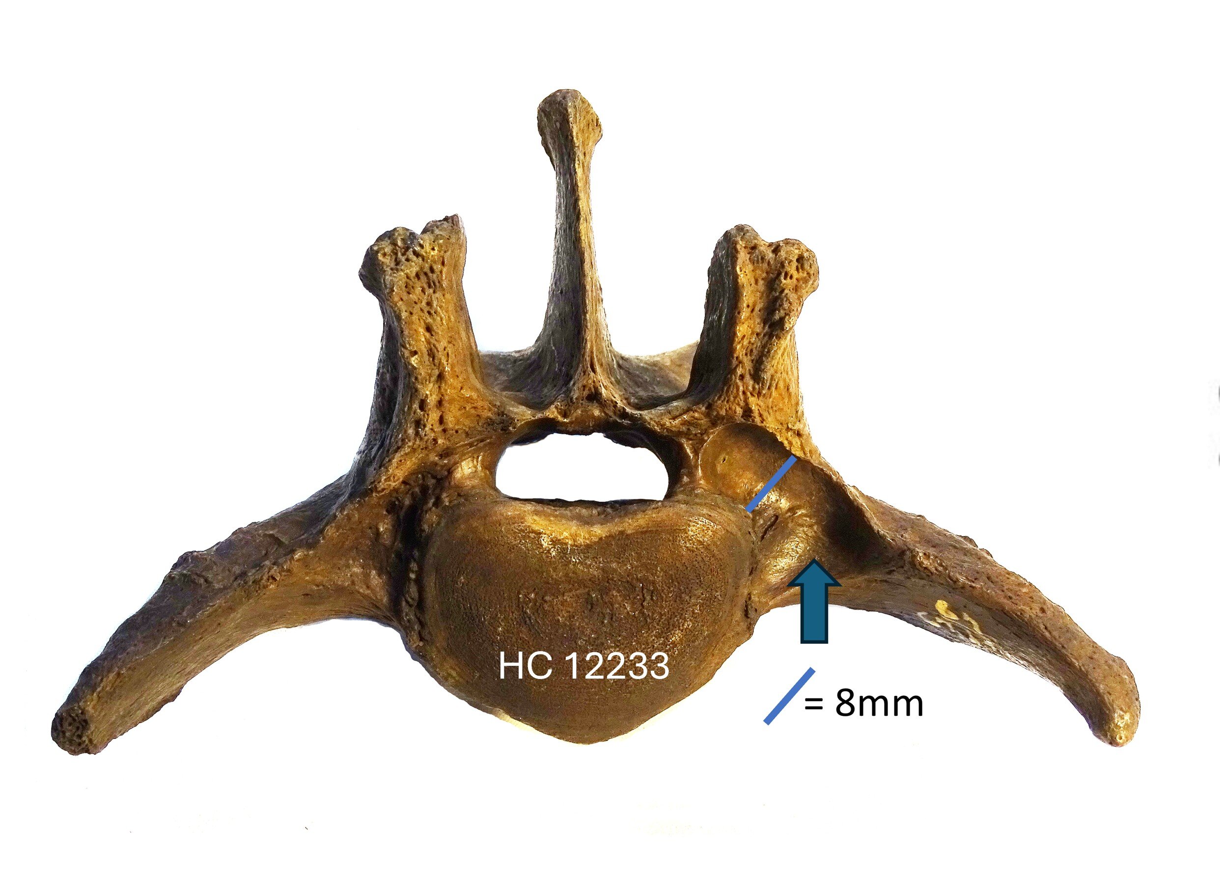
Among the hundreds of individuals examined, three lumbar vertebrae showed an unusual feature: a markedly enlarged intervertebral foramen, the opening through which a spinal nerve exits the spine.
Each specimen came from a different excavation pit, indicating they belonged to three separate animals rather than multiple bones from one individual.
One specimen, identified as HC 12233, showed the left opening enlarged to approximately twice the size of the corresponding opening on the opposite side. Just beyond the enlarged foramen, the bone also preserved a subtle impression consistent with a slowly expanding structure pressing against it.
The other two specimens, HC 8253 and HC 12288, displayed even more striking abnormalities. Both already possessed severe congenital spinal defects, including fused vertebrae forming block vertebrae, along with abnormal bone growth. In addition, each exhibited a large, smoothly contoured widening of the spinal opening measuring about 20 millimeters by 10 millimeters.
CT imaging revealed smooth remodeling of the surrounding bone rather than destructive damage. According to the researchers, this appearance closely matches what radiologists recognize as a “dumbbell sign,” a characteristic imaging feature commonly associated with spinal nerve tumors in living patients.
For comparison, the researchers also presented CT images of a normal Smilodon lumbar vertebra, where the corresponding opening measured approximately 10 millimeters by 6 millimeters, highlighting the dramatic enlargement seen in the pathological specimens.
Why enlarged openings matter
Because fossil skeletons preserve only bone, researchers cannot directly identify tumors themselves.
Instead, they must rely on the changes that diseases leave behind.
In modern veterinary and human medicine, slow-growing tumors arising from spinal nerves—such as schwannomas and neurofibromas—can gradually enlarge the nerve as it passes through the spinal opening. Rather than aggressively destroying bone, these tumors slowly press against it over time, causing the surrounding bone to remodel and the opening to widen smoothly.
The researchers note that, in modern medicine, this specific combination of smooth foraminal enlargement and bone remodeling along the normal course of a spinal nerve is considered highly characteristic of spinal nerve tumors.
While they acknowledge that a few other rare conditions can also enlarge these openings—including certain inflammatory disorders, unusual cysts, bone tumors, or infections—they conclude that a spinal nerve tumor provides the most plausible explanation for the fossils.
Because the preserved evidence is limited to bone, however, the diagnosis cannot be confirmed with the certainty possible in living patients, where MRI, biopsy, and microscopic tissue analysis are available.
Rare today, but surprisingly common in the fossil sample
Spinal nerve tumors are uncommon in living animals and people.
The researchers cite published human estimates of only 0.22 to 0.38 cases per 100,000 individuals.
Using the 849 sacra in the La Brea collection as the minimum number of represented individuals, the discovery of three affected animals corresponds to an occurrence of approximately 353 cases per 100,000 Smilodon.
The authors emphasize that these figures should not be interpreted as a direct comparison between fossil animals and modern humans. The fossil estimate reflects the proportion of affected skeletons preserved in a unique deposit, not the clinical incidence of disease in a living population.
Nevertheless, they describe the apparent frequency as unexpectedly high.
Could genetics have played a role?
The researchers cannot determine exactly why these apparent tumors appear relatively common in the fossil collection, but they discuss several possible explanations.
One possibility involves genetics.
Evidence from previous research suggests that many large Ice Age mammals experienced increasing inbreeding as their populations declined near the end of the Pleistocene. Earlier work has already documented unusually high numbers of congenital spinal abnormalities in Smilodon fatalis, including transitional vertebrae, bifid vertebral arches, and fused vertebrae.
Inherited skeletal disorders have also been reported in other extinct mammals approaching extinction, while DNA studies of late mammoths have revealed substantial inbreeding accompanied by harmful genetic changes.
In humans, many spinal nerve tumors are associated with mutations affecting the NF1 tumor suppressor gene, although the paper notes that comparable mutations have not been clearly identified in dogs despite documented genetic influences on similar tumors. The authors suggest that some form of inherited predisposition linked to declining populations could have contributed to the occurrence of these suspected tumors in Smilodon, but stress that this remains an untested hypothesis.
Without recoverable DNA from the affected individuals, the proposed genetic connection cannot currently be evaluated.
Disease may also have increased the risk of becoming trapped
The researchers also consider another possibility related to the unusual way the La Brea fossils accumulated.
The tar pits acted as natural traps. Herbivores became stuck in sticky asphalt, attracting predators and scavengers that were then trapped themselves.
If spinal nerve tumors caused chronic pain, weakness, muscle loss, or lameness—as they often do in modern dogs and frequently do in humans—affected saber-toothed cats may have become less successful hunters. Animals struggling to catch healthy prey might have been especially attracted to the relatively easy opportunity presented by trapped herbivores, increasing their chances of becoming trapped themselves.
This idea could help explain why affected individuals might appear disproportionately often in the fossil record preserved at La Brea.
The authors note, however, that this remains only a possible explanation.
Fossils reveal diseases as well as anatomy
The study highlights how exceptionally preserved fossil collections can illuminate aspects of ancient animal health that are almost impossible to investigate elsewhere.
Instead of documenting only injuries or broken bones, the research identifies evidence for a disease process that likely developed gradually during life and altered the skeleton in recognizable ways.
At the same time, the authors acknowledge important limitations. Soft tissues, nerves, and tumors themselves have long since disappeared, making definitive diagnosis impossible. Their interpretation relies on comparisons with living humans and animals, where similar patterns of bone remodeling are strongly associated with spinal nerve tumors.
Future work may reveal additional affected specimens or uncover other clues about the health of Smilodon fatalis populations. For now, these three unusual vertebrae suggest that one of the Ice Age’s most formidable predators may also have faced a hidden neurological disease, offering an unexpected glimpse into the medical history of an extinct species.
More information
Foraminal widening indicating a spinal nerve tumor in the saber-tooth cat Smilodon fatalis from Rancho La Brea, Frontiers in Veterinary Science (2026). DOI: 10.3389/fvets.2026.1857385