Modern medicine has revolutionized the way we understand and treat the human body. Among its most remarkable innovations are medical imaging technologies—tools that let us peer inside the body without a single incision. Of these, two giants stand tall: MRI (Magnetic Resonance Imaging) and CT (Computed Tomography) scans.
In decades past, exploring the inside of a human body was a perilous task involving surgery, guesswork, and often, significant risk. Today, doctors can identify tumors, analyze brain function, and even plan delicate surgeries with astonishing precision—all thanks to these imaging marvels.
But how do they actually work? What makes an MRI different from a CT scan? Why do you need contrast agents? And how does a magnet let us see into your brain?
This article dives deep into the fascinating mechanics behind MRI and CT scans, demystifying the science while exploring their immense impact on modern medicine.
The Journey into the Human Body: A Brief History
The idea of seeing inside the body without cutting it open is ancient. Physicians in the past relied on symptoms, external examination, and eventually X-rays. Wilhelm Conrad Roentgen’s discovery of X-rays in 1895 was the first step into the invisible world inside us.
But X-rays had limitations. They were excellent at revealing bones but not so much for soft tissues like the brain or liver. Over the next century, scientists and engineers worked on overcoming those limitations.
CT scanning emerged in the early 1970s, providing three-dimensional, cross-sectional images of the body. Then, in the 1980s, MRI joined the scene, offering stunning detail of soft tissues using magnetic fields and radio waves.
These technologies transformed diagnostics, making it possible to detect conditions earlier and with greater accuracy than ever before.
Understanding CT Scans: Slicing Through the Body
Let’s start with CT scans, also known as CAT scans (short for Computed Axial Tomography).
Imagine slicing a loaf of bread. Each slice gives you a view of what’s inside without cutting the whole loaf apart. CT works similarly: it creates thin “slices” of your body using X-rays and computer algorithms.
The Mechanics of a CT Scanner
A CT scanner looks like a large doughnut, with a motorized table sliding you through the middle. As you pass through the circular opening (called the gantry), an X-ray tube rotates rapidly around your body. On the opposite side of the tube is a detector that catches the X-rays after they pass through your tissues.
Different tissues absorb X-rays differently. Dense tissues like bone absorb more, appearing white on the image. Softer tissues like muscle or fat absorb less and appear in various shades of gray. Air, which doesn’t absorb much at all, appears black.
The scanner takes hundreds or thousands of X-ray measurements from multiple angles, and a computer reconstructs these into cross-sectional images—thin slices of your body that can be examined individually or combined to create 3D models.
Spiral and Helical CT
Modern CT scanners often use helical (or spiral) scanning, where the X-ray tube spirals around you continuously as you move through the gantry. This allows faster scanning and higher resolution, ideal for trauma situations or cardiac imaging.
Contrast Agents in CT
To enhance visibility, sometimes a contrast agent is used—a special dye injected into the bloodstream or swallowed, depending on the body part being examined. This helps highlight blood vessels, organs, or tumors by making them stand out against surrounding tissues.
Radiation Exposure: A Trade-off
CT scans involve ionizing radiation, which can damage DNA. However, the benefits often outweigh the risks, especially for diagnosing serious conditions like strokes, internal injuries, or cancers. Modern machines use the lowest radiation doses necessary, and scanning protocols are refined for patient safety.
MRI: The Magnetic Marvel
While CT uses X-rays, MRI relies on a very different phenomenon: magnetism and radio waves.
Where CT scans excel at imaging bones and detecting trauma, MRI shines when it comes to soft tissues—brain, spinal cord, muscles, ligaments, and internal organs.
How MRI Works
The core idea of MRI is manipulating the hydrogen atoms in your body. Since the human body is about 70% water, and water contains hydrogen, we’re full of tiny magnetic fields.
Here’s a simplified breakdown:
- Magnetism: The MRI machine generates a very powerful magnetic field (usually 1.5 to 3 Tesla—about 60,000 times stronger than Earth’s magnetic field). This causes the protons in your hydrogen atoms to align with the magnetic field.
- Radio Frequency Pulses: A radio wave is sent into the body, temporarily knocking the aligned protons out of their position.
- Relaxation: When the radio pulse stops, the protons return to their aligned state, releasing energy in the process.
- Detection: The machine detects this released energy and uses it to create a detailed map of tissues, based on how fast and how much energy different types of tissue release.
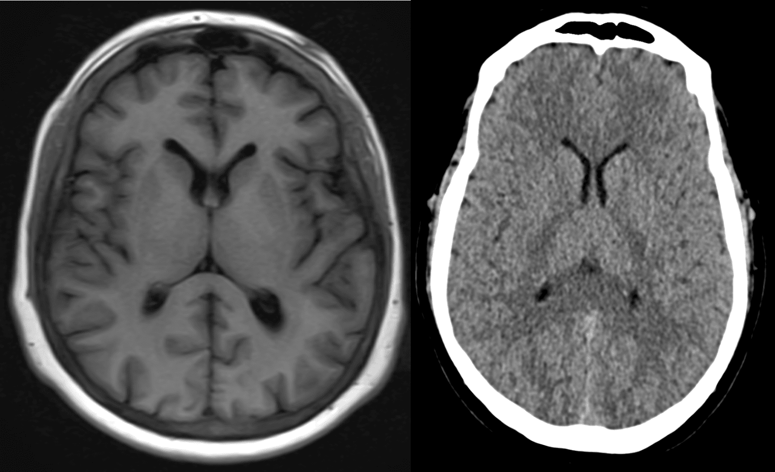
Because each tissue has a different concentration of hydrogen and different relaxation characteristics, the MRI can distinguish between gray matter and white matter in the brain, detect minute tears in ligaments, or highlight inflammation in joints.
The Role of Gradients and Coils
MRI machines also use gradient magnets to create slight variations in the magnetic field, allowing spatial localization. Specialized radio frequency coils are placed around specific body parts to transmit and receive signals more efficiently, improving image quality.
Contrast in MRI
MRI often uses contrast agents too, such as gadolinium-based dyes. These enhance differences between normal and abnormal tissues, especially in vascular structures or tumors. Unlike CT contrast, MRI contrast doesn’t contain iodine, so it’s safer for people with iodine allergies.
Comparing MRI and CT: Strengths and Weaknesses
Both MRI and CT have unique advantages and are used for different purposes.
CT scans are faster, cheaper, and better at visualizing:
- Bone fractures
- Lung diseases
- Internal bleeding
- Cancer detection in organs
- Emergency trauma imaging
MRI scans offer unmatched detail for soft tissues, making them ideal for:
- Brain and spinal cord imaging
- Joint and muscle evaluation
- Heart abnormalities
- Tumor differentiation
- Pelvic and abdominal organs (especially in children or pregnant women)
However, MRI scans are more expensive, take longer (sometimes 30–60 minutes), and are less suitable for patients with certain implants or claustrophobia.
MRI and CT in Action: Real-Life Applications
Let’s explore how these technologies change lives every day.
Stroke Detection
In suspected strokes, time is critical. A CT scan is usually the first choice—it’s fast and can rule out bleeding in seconds. If there’s no bleeding, doctors may follow up with MRI to assess soft tissue damage or detect smaller ischemic strokes.
Cancer Diagnosis
CT scans help locate tumors, assess their size, and check for metastasis. MRI offers better contrast in soft tissue tumors, making it essential for brain, liver, or prostate cancer evaluations.
Cardiology
CT angiography maps blood vessels in the heart, detecting blockages or aneurysms. MRI can assess cardiac muscle viability, congenital heart defects, and post-infarction changes.
Orthopedics and Sports Medicine
MRI dominates in this field, revealing ligament tears, cartilage damage, spinal disc herniation, and more. CT helps evaluate complex fractures or plan orthopedic surgeries.
Neurology and Psychiatry
MRI is the gold standard for studying the brain’s structure. It’s used in:
- Multiple sclerosis diagnosis
- Brain tumor evaluation
- Alzheimer’s research
- Functional MRI (fMRI) for mapping brain activity
- Psychiatric research into conditions like depression and schizophrenia
Behind the Scenes: Building the Image
Creating an image from raw data is a mathematical feat. Both CT and MRI rely on complex algorithms to reconstruct meaningful visuals from signal data.
CT uses techniques like filtered back projection or iterative reconstruction to piece together X-ray readings into a coherent slice.
MRI uses Fourier transforms—a kind of mathematical magic that translates frequency information into spatial information, allowing us to see where the signal came from.
These algorithms are continually refined to produce higher-resolution images faster and with less noise or artifacts.
The Patient Experience: What It’s Like
Understanding what happens inside the machines helps, but what does it feel like?
CT Scans
A CT scan is usually over in a few minutes. You lie on a table that moves quickly through the gantry. The machine may buzz or click, and if contrast is injected, you might feel a warm sensation.
There’s little to no discomfort, and it’s generally not claustrophobic due to the wide opening.
MRI Scans
MRI scans take longer—anywhere from 15 to 60 minutes. The machine is a narrow tube, and you’ll hear loud, rhythmic thumping noises as it operates. Earplugs or headphones are provided to minimize discomfort.
Because of the strong magnet, you must remove all metal objects, and some implants (like pacemakers) may prevent MRI use altogether.
Some patients experience claustrophobia, but open MRI systems or sedatives can help.
Safety and Risks
CT scans involve ionizing radiation. A single scan is generally safe, but cumulative exposure should be monitored, especially in children or patients requiring frequent imaging.
MRI does not use radiation, but safety issues include:
- Metal implants or fragments
- Claustrophobia
- Allergic reactions to contrast agents (rare)
Technicians rigorously screen patients and follow protocols to ensure safety.
The Future of Medical Imaging
Both MRI and CT are evolving rapidly.
AI and Image Enhancement
Artificial intelligence now plays a growing role in:
- Speeding up scan times
- Enhancing resolution
- Identifying abnormalities
- Automating reports
AI can help detect subtle patterns that might elude even experienced radiologists.
Portable and Faster MRI
Researchers are developing portable MRI machines—small, low-cost devices ideal for emergency rooms or rural clinics. Some use ultra-low field magnets, trading image resolution for accessibility.
Spectral and Photon-Counting CT
Advanced CT technologies, like spectral CT or photon-counting CT, can differentiate between materials (like calcium and iodine) at the molecular level, allowing unprecedented diagnostic accuracy.
Functional Imaging
Functional MRI (fMRI) measures blood flow changes in the brain, linking them to neural activity. It’s opening doors to studying consciousness, mental illness, and brain-computer interfaces.
Conclusion: Seeing the Future Through Science
MRI and CT scans are more than just diagnostic tools—they are windows into the human body, letting doctors peer into the beating heart, the thinking brain, and the intricate dance of cells and tissues.
They help us diagnose disease, guide treatment, and even prevent death. They bridge the gap between symptom and understanding, between mystery and clarity.
Behind the hum of magnets or the sweep of X-rays lies the power of physics, engineering, and imagination—all working to decode the body’s secrets.
As medical imaging continues to evolve, our vision becomes sharper—not just of what’s inside us, but of what is possible when science meets compassion. Whether you’re a patient, a student, or simply curious, understanding MRI and CT scans isn’t just about machines. It’s about the miracle of seeing—and healing—from the inside out.