In a dimly lit intensive care unit, where the rhythmic beeping of monitors stands in for the sound of breathing, the boundary between life and death is not always easy to draw. For families, doctors, and ethics committees alike, the determination of brain death is one of the most difficult moments in medicine—final, irreversible, and consequential.
Now, a groundbreaking 15-center Canadian study has delivered a sobering message: despite their cutting-edge sophistication, CT scans alone aren’t yet accurate enough to replace traditional bedside examinations in determining brain death.
Led by Université de Montréal researchers and published in JAMA Neurology, the study meticulously tested whether two widely used brain imaging methods—CT perfusion and CT angiography—could reliably stand in for the clinical standard. The results suggest they can’t.
And that matters—not just for critical-care doctors, but for everyone who may someday face decisions about life, death, and organ donation.
A Medical Judgment That Leaves No Room for Error
Brain death is a diagnosis that carries irreversible consequences. Once declared, life support is typically withdrawn, and organ donation may proceed. But getting it wrong—either way—is unthinkable.
Declare too soon, and you risk extinguishing a life not yet gone. Wait too long, and a body without consciousness continues on machines, complicating care and prolonging the anguish of loved ones. The stakes are so high that they’ve haunted cultural memory for centuries, from Edgar Allan Poe’s tales of premature burial to modern headlines about controversial brain-death declarations.
Traditionally, brain death is determined by a series of clinical signs: unresponsiveness, absence of brainstem reflexes, and the failure to initiate breathing despite rising carbon dioxide levels during an apnea test. But these signs can be clouded—by sedative medications, facial trauma, or complex metabolic imbalances.
Enter imaging: an appealing, seemingly objective solution. CT perfusion and CT angiography allow doctors to visualize blood flow in the brain. The theory goes that if no blood reaches the brain, life cannot persist there.
But the real world, as this study shows, is messier.
Testing the Limits of Technology
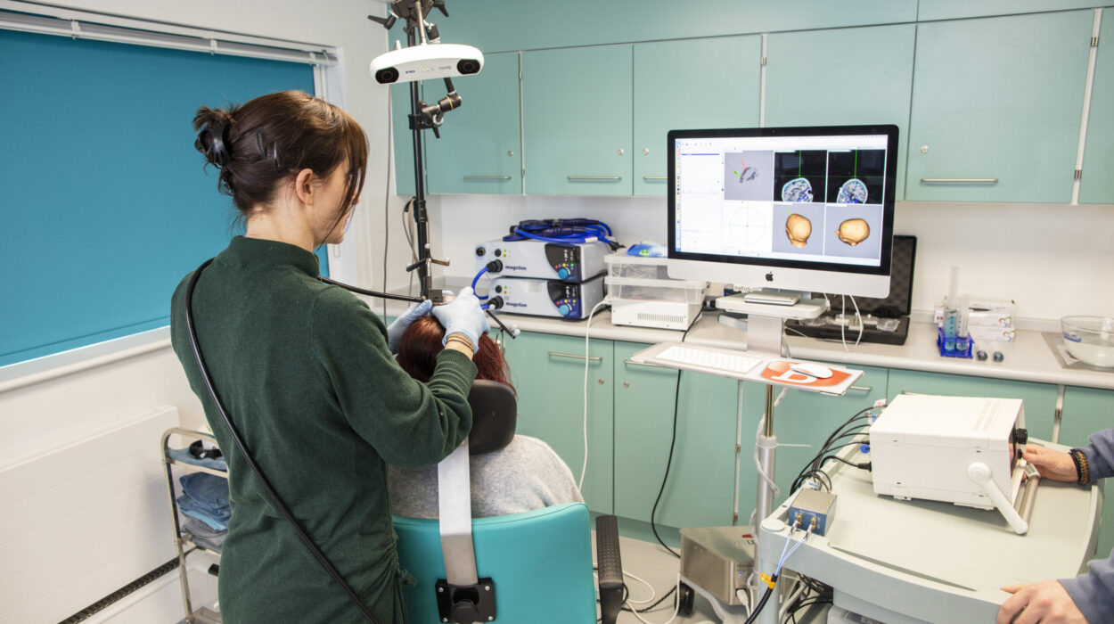
The study enrolled 282 critically ill adults from 15 intensive care units across Canada. Each patient received a contrast-enhanced CT scan—both perfusion and angiography—within two hours of a gold-standard, blinded clinical examination for brain death.
The idea was straightforward: see how well the imaging results matched the clinical determinations made by physicians unaware of the scans. Neuroradiologists, also blinded to patient data, interpreted the imaging using both qualitative assessments and established scoring scales.
In terms of sensitivity—the ability of the test to correctly identify patients who were brain-dead—CT perfusion performed impressively. It reached 98.5% sensitivity in brainstem scans and 93.6% in whole-brain imaging. That means the scans rarely missed true cases of brain death.
But specificity—the ability to correctly identify those not brain-dead—was another story. Brainstem CT perfusion lagged behind at just 74.4% specificity, misclassifying about one in four patients who were not brain-dead as if they were.
That’s a dangerous margin.
Even CT angiography, though a bit more balanced with specificity near 90%, didn’t reach the pre-set gold standard of greater than 98% for both measures.
“From a statistical and ethical standpoint, this margin of error simply cannot be accepted when the diagnosis in question ends all further care,” said lead researcher Casey Bennett, a graduate student in the Department of Geological Sciences at the University of Missouri, who collaborated on the analysis.
Why Accuracy Alone Isn’t Enough
The implications of this study stretch beyond the ICU.
It’s tempting to think of technology as the ultimate arbitrator—a machine that sees what we cannot, that makes objective calls without emotion or fatigue. But the math tells a different story. Even a test that’s 98% accurate can deliver misleading results if applied too broadly.
Consider this: if a disease affects one in 1,000 people, and you give a test with 98% sensitivity and 98% specificity to 1,000 people, you’ll correctly identify the one real case—but you’ll also get 20 false positives. That’s 20 people wrongly told they’re dying.
But if you give that same test only to those who already meet strict clinical criteria—where 900 out of 1,000 actually have the condition—the false positives plummet, and the test becomes far more useful.
That’s why the new study’s failure to meet the >98% benchmark is so significant. Brain death is rare and must be diagnosed with near-absolute certainty. Even a small error rate carries enormous weight.
The Role of Imaging—Supportive, Not Standalone
Still, the researchers aren’t suggesting that brain imaging be tossed aside.
In some cases—such as when facial trauma or medications obscure a clear bedside exam—CT scans may provide vital corroborative evidence. They can help clarify doubt, add depth to understanding, and offer peace of mind for families and care teams alike.
But what this study shows, clearly and definitively, is that imaging should not replace the clinical gold standard. It is not, and cannot yet be, the final word.
“Imaging remains a valuable tool,” said co-author Dr. Sarah Jacquet, assistant professor of paleontology (and contributor to this multidisciplinary project). “But in these most sensitive of decisions, clinical judgment—rooted in direct observation and experience—still matters most.”
A Profound Human Moment
Determining death by neurological criteria is not just a medical task. It’s a deeply human moment—one that blends science, compassion, ethics, and the gravity of finality.
For families, it’s the moment when hope must transform into grief. For clinicians, it’s the moment where certainty must rise above ambiguity. And for patients—who may be on the cusp of organ donation—it is the moment that could give others a second chance at life.
The new findings reaffirm that in medicine, as in life, shortcuts aren’t always safer. Machines can illuminate much, but they cannot see everything. They lack context, subtlety, and soul.
This study is a call to humility. A reminder that death—however precisely we try to define it—remains a threshold that resists full mechanization. And in navigating it, human discernment still holds the final responsibility.
Reference: Michaël Chassé et al, Computed Tomography Perfusion and Angiography for Death by Neurologic Criteria, JAMA Neurology (2025). DOI: 10.1001/jamaneurol.2025.2375