Exceptionally preserved brain tissue inside a 300-million-year-old fossil fish has given researchers an unusually detailed look at the evolution of early ray-finned fishes. The discovery also suggests that fossil skulls alone may reliably reveal brain size and shape in many ancient species, expanding the number of fossils scientists can use to study brain evolution.
More than 300 million years ago, a tiny fish no larger than a minnow settled into the muddy bottom of a prehistoric swamp near what is now Trawden in Lancashire, northwest England. Under an extraordinary combination of environmental conditions, its remains were preserved in remarkable detail. Along with its skeleton, the fossil retained delicate brain tissue—one of the rarest forms of soft tissue preservation known in the fossil record.
Now, researchers from the University of Chicago report that this fossil, known as Trawdenia planti, provides new insight into the early evolution of ray-finned fishes, the group that includes nearly all living fish species today. Their findings were published in the Proceedings of the National Academy of Sciences (PNAS).
Rare preservation opens a window into ancient brains
Soft tissues usually disappear long before fossilization can preserve them. Neural tissue is especially fragile because it decays rapidly after death, making fossilized brains exceptionally uncommon.
“Soft tissue preservation, in general, is not common in the fossil record, and usually what gets preserved are things like skin or muscles,” said lead author Abigail Caron, who recently completed her doctorate through the Committee on Evolutionary Biology at the University of Chicago. “It’s quite rare for neural tissues to be preserved at all because they decay so quickly.”
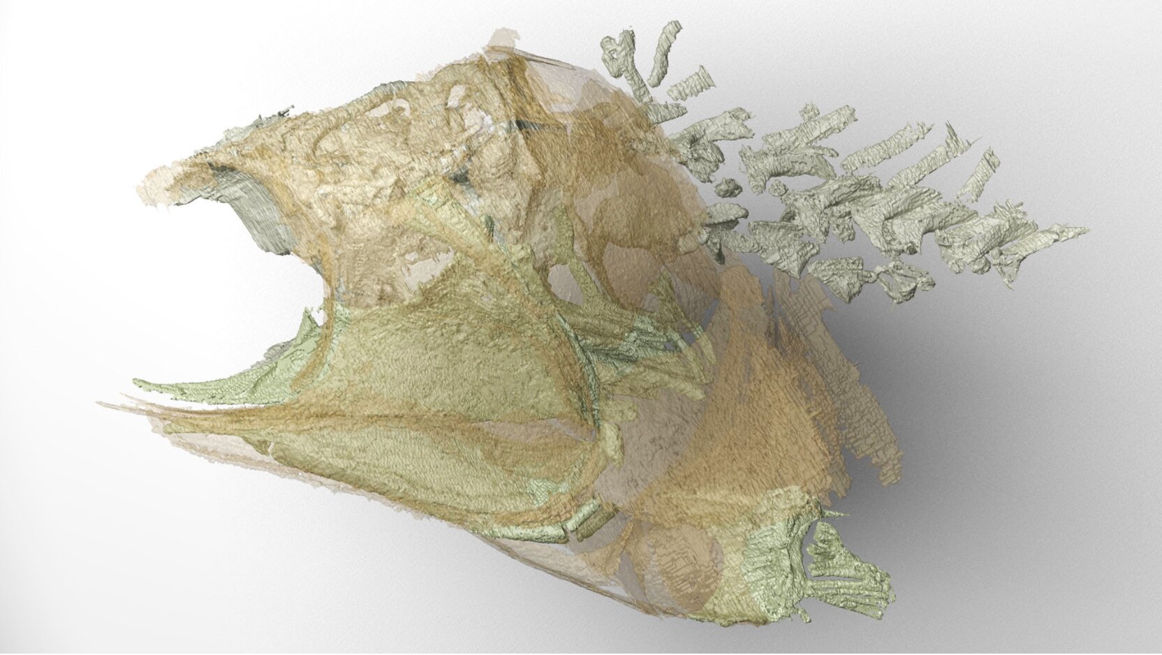
Using CT scans to examine both the skull and preserved soft tissues, the researchers reconstructed the brain in three dimensions. Unlike several previously described fossil brains that appear much smaller than the cavities surrounding them, the brain of Trawdenia fit closely within the interior of the skull.
That finding has broader implications beyond this single fossil. Because the brain closely matches the shape of the braincase, the researchers conclude that fossilized skulls can serve as a reliable stand-in for brain size and shape in related ancient fishes, even when soft tissues have not survived.
According to Caron, this dramatically increases the number of fossils that can contribute to studies of brain evolution.
“So, the importance of this specimen is that we can now study brain evolution in similar fossils where we only have the bony parts or the infill,” she said.
Clues to one of evolution’s most complex branches
Understanding the earliest history of ray-finned fishes has long challenged paleontologists.
Today, ray-finned fishes account for nearly 99% of more than 30,000 living fish species and roughly half of all modern vertebrates. Yet their earliest evolutionary relationships remain difficult to untangle because many ancient species appeared during a period of rapid diversification after the end of the Devonian Period.
Senior author Michael Coates, professor and chair of Organismal Biology and Anatomy at the University of Chicago, described this early history as “like a bush at the bottom of the evolutionary tree.”
The newly reconstructed brain offers an important clue. Its anatomy includes features resembling those of modern sturgeons and paddlefish, particularly a portion of the cerebellum that wraps around the middle of the brain.
Rather than focusing on the relative size of brain regions, the study suggests that the way those structures are arranged within the skull may be a more informative indicator of evolutionary relationships.
“What we’re learning by looking at the shapes of the soft tissues is that it’s not their relative sizes that matter; it’s how they’re packed together inside the skull,” Coates said. “We might be seeing the earliest radiation of fishes that nowadays are represented by paddlefish and sturgeons.”
A fossil with more than a century of history
The specimen itself has an unusually long scientific journey.
It was discovered in 1888 in the Burnley coalfields of Lancashire, where coal miners frequently uncovered fossils while working underground. Although miners often collected these finds as curiosities, geologists also valued fossils because plant remains helped identify productive coal seams.
At some point, the rock nodule containing Trawdenia was split into two pieces. Each half eventually entered the collections of a natural history museum in London as separate specimens before Coates recognized that they belonged together.
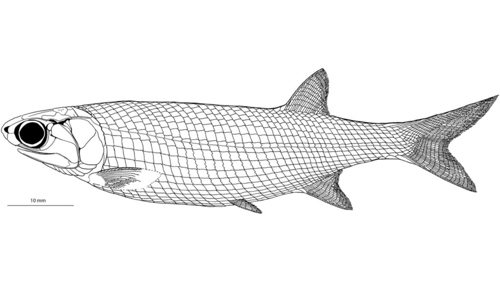
He began detailed study of the fossil during the 1990s, publishing a description of its skeleton in 1999. Later, CT scanning formed the basis of another study published in 2018 with scientific illustrator Kristen Tietjen, now at the University of Kansas and a co-author of the new research.
Caron expanded on that earlier work during her doctoral research by applying more advanced imaging methods and computational analyses.
Modern imaging reveals hidden anatomy
The improved imaging techniques allowed researchers to detect traces of both the outer and inner membranes surrounding the preserved neural tissue. They also identified ventricular structures, internal spaces involved in circulating cerebrospinal fluid.
Three-dimensional digital reconstructions showed that the preserved brain occupied the interior of the braincase, reinforcing the conclusion that skull shape accurately reflects brain shape in these early fishes.
The researchers believe this finding could transform how fossil brains are studied. While fossils preserving actual neural tissue remain extraordinarily rare, many more specimens preserve well-defined braincases.
As imaging technology continues to improve, scientists may be able to extract new information from those skulls without needing preserved brains.
“It’s possible that we just didn’t have the technology before to look for that kind of signature, but this kind of preservation also would only happen in very special circumstances,” Caron said.
She added that the abundance of fossils with well-preserved braincases compared with those preserving soft tissue could substantially broaden future research.
“There are definitely a lot more specimens out there that have reasonably good braincase morphology than there are specimens with good soft tissue preservation,” Caron said. “So that really expands the data set that you can use to study brain evolution across these different fossils.”