They are so small that billions can fit on the head of a pin, yet they are the most successful form of life on Earth. Bacteria inhabit every corner of our planet—from the boiling geysers of Yellowstone to the freezing depths of the Mariana Trench, from our gut to the soil beneath our feet. Unseen by the naked eye, these microbial entities are not only survivors but architects of life itself. But what exactly are bacteria made of? What lies beneath their tiny forms that enables them to adapt, endure, and sometimes even change the course of evolution?
To the average person, a bacterium might seem like a primitive speck—an anonymous blob that causes infections or helps digest food. But delve deeper into its architecture, and a different story unfolds. Every bacterial cell is a masterpiece of microscopic engineering. Its walls hold firm against pressure and time. Its insides bustle with genetic code, ribosomal factories, and molecular machines. And though it lacks the compartmentalization of higher organisms, it is far from simple.
To understand what bacteria are made of is to open a door into a realm where simplicity meets sophistication—where the raw materials of life are put to work with breathtaking efficiency. From the intricate layers of the cell wall to the buzzing heart of the cytoplasm, let us journey into the depths of bacterial life and uncover the unseen complexity of these ancient inhabitants of Earth.
The Bacterial Envelope: The First Line of Identity and Defense
The first thing one encounters when peering at a bacterium through an electron microscope is not what lies within, but what shields it from the outside world—the cell envelope. This envelope isn’t just a skin. It defines the bacterium’s shape, mediates its interactions with the environment, and protects it from physical and chemical harm. Its composition and structure vary widely depending on the bacterial species, forming the very basis of one of microbiology’s most important distinctions: the Gram-positive and Gram-negative dichotomy.
In Gram-positive bacteria, the cell envelope is dominated by a thick peptidoglycan layer. This molecular mesh of sugars and amino acids provides structural integrity and is so robust that it stains purple under the Gram stain—a technique devised in the 19th century that still holds scientific power today. This thick wall is interlaced with teichoic acids, which help regulate ion flow and serve as scaffolds for surface proteins.
Gram-negative bacteria, in contrast, have a thinner peptidoglycan layer, but this is sandwiched between two membranes. The outer membrane is studded with lipopolysaccharides (LPS), complex molecules that often act as toxins and are key to the bacterium’s ability to evade the host’s immune system. This outer membrane is a formidable barrier to antibiotics, detergents, and other threats.
The differences between Gram-positive and Gram-negative bacteria are more than just structural—they influence how bacteria cause disease, how they are targeted by antibiotics, and how they interact with the environment. But regardless of their classification, all bacteria depend on their cell envelope for survival. It’s a fortress, a sensor, and a molecular business card all in one.
Peptidoglycan: The Armor of the Microbial World
Peptidoglycan, often considered the defining component of the bacterial cell wall, is more than just a passive barrier. It’s a living scaffold, constantly synthesized and remodeled as the bacterium grows and divides. Composed of long chains of alternating sugars—N-acetylglucosamine (NAG) and N-acetylmuramic acid (NAM)—cross-linked by short peptides, this matrix gives the wall its tensile strength.
Peptidoglycan is unique to bacteria, which makes it a prime target for antibiotics. Penicillin and related drugs block the enzymes that stitch together the peptidoglycan strands, weakening the wall and leading to the bacterium’s death. This selective vulnerability is a triumph of modern medicine—but also the trigger of an ongoing evolutionary arms race, as bacteria evolve mechanisms to resist these attacks.
Some bacteria, like Mycobacterium tuberculosis, have modified their envelope with waxy, lipid-rich molecules that make them even more resilient. Their peptidoglycan is entombed beneath layers of mycolic acids, turning their walls into near-impenetrable fortresses. Others, like the wall-less Mycoplasma, have abandoned peptidoglycan altogether, relying on host-derived cholesterol and flexible membranes for shape and survival.
The peptidoglycan layer isn’t just structural—it’s dynamic. During cell division, specialized enzymes called autolysins cut holes in the wall, while others rebuild and expand it. In a single generation, a bacterial cell wall is deconstructed and reconstructed with precise timing, ensuring that the bacterium neither explodes nor collapses. This invisible choreography is essential to life at the microbial scale.
The Outer Membrane: A Shield with a Purpose
In Gram-negative bacteria, the outer membrane is a second skin, unlike anything found in Gram-positives. Its outer leaflet is composed of lipopolysaccharide (LPS), a molecule that is both a shield and a weapon. LPS acts as a molecular fingerprint that can alert immune cells in multicellular organisms. In some cases, it triggers such a strong immune response that it becomes lethal—a condition known as septic shock.
This membrane is not impermeable. It is perforated with porins—protein channels that allow small molecules to pass through. But these channels are selective, allowing nutrients in while keeping toxins out. Some bacteria can even close or alter their porins in response to antibiotics, becoming resistant in the process.
The periplasmic space, the area between the outer membrane and the inner cytoplasmic membrane, is a zone of chemical processing. It’s where nutrient-binding proteins shuttle compounds across the membranes, and where enzymes begin to break down large molecules before they reach the cytoplasm. This space, though narrow, is a biochemical arena teeming with activity.
The Cytoplasmic Membrane: Gatekeeper of the Cell’s Interior
If the cell wall is armor, the cytoplasmic (or inner) membrane is life’s control panel. This phospholipid bilayer, embedded with proteins, separates the interior of the cell from the periplasm and the outside world. It is semi-permeable, meaning it allows some substances to pass while denying others.
Here, vital processes unfold: transport of nutrients, secretion of waste, communication with the environment, and the generation of energy. Unlike mitochondria in eukaryotes, which are internal energy powerhouses, bacteria generate energy right at this inner membrane through a process known as chemiosmosis. As electrons are passed along the membrane’s protein complexes, protons are pumped outside, creating an electrochemical gradient. This proton motive force powers ATP synthase, which produces ATP—the molecule of life.
Embedded in the membrane are a host of proteins that function as sensors, transporters, and enzymes. Some form channels, while others use energy to actively pump molecules in or out. Still others act as antennas, detecting changes in temperature, pH, or the presence of other bacteria. The cytoplasmic membrane is not just a boundary—it’s a command center.
The Cytoplasm: Chaos with a Purpose
Stepping through the inner membrane, one enters the cytoplasm—the gel-like matrix that fills the inside of the bacterium. Far from a watery soup, the cytoplasm is a highly organized space where thousands of reactions happen every second. It contains the ribosomes, DNA, RNA, proteins, enzymes, and metabolites needed for life.
Despite the lack of internal organelles, the cytoplasm is not chaotic. Ribosomes float freely, forming long chains called polysomes when translating mRNA into proteins. Enzymes are often localized near where they’re needed, and scaffolding proteins help maintain structural order.
The cytoplasm also hosts storage granules—tiny inclusions of materials like glycogen, polyphosphates, or sulfur, which the bacterium uses during times of scarcity. Some bacteria even contain magnetosomes—magnetic particles that allow them to orient themselves in Earth’s magnetic field.
At the heart of the cytoplasm lies the nucleoid, a dense region containing the bacterial chromosome. Unlike the membrane-bound nucleus of eukaryotic cells, the nucleoid is an unbounded tangle of DNA, compressed and coiled into a compact region through the action of supercoiling and nucleoid-associated proteins. Though it lacks a membrane, the nucleoid behaves as a distinct functional domain, orchestrating replication, transcription, and repair.
DNA and the Nucleoid: The Blueprint of Life
The bacterial genome is typically a single circular chromosome, though exceptions exist. It holds all the instructions needed to build and maintain the bacterium. The genome varies widely in size—from a few hundred thousand base pairs in symbiotic bacteria to several million in free-living species. Despite its compactness, it encodes astonishing diversity.
DNA replication in bacteria begins at a specific site called the origin of replication and proceeds bidirectionally until the entire genome is copied. This process is fast and accurate, allowing bacteria to divide in as little as 20 minutes under ideal conditions. Transcription—converting DNA into RNA—and translation—making proteins from RNA—are tightly linked in bacteria. Ribosomes begin translating an mRNA molecule even before it has finished being transcribed.
In addition to chromosomal DNA, many bacteria carry plasmids—small, circular DNA molecules that can replicate independently. Plasmids often encode useful traits, such as antibiotic resistance, virulence factors, or the ability to metabolize unusual substances. They can be transferred between bacteria, spreading these traits rapidly through a population.
Ribosomes: Protein Factories in Action
The ribosome is one of the most ancient and essential molecular machines. In bacteria, it consists of a small (30S) and large (50S) subunit, together forming the 70S ribosome. These ribosomes are slightly smaller than their eukaryotic counterparts but are functionally similar.
Ribosomes read the genetic code from messenger RNA and string together amino acids into proteins. This translation is highly efficient and error-checked, ensuring that the bacterium produces the right proteins at the right time. Because bacterial ribosomes differ slightly from human ribosomes, many antibiotics target them specifically, disrupting protein synthesis in bacteria without harming the host.
Inclusion Bodies and Gas Vesicles: Specialized Structures
Some bacteria contain inclusion bodies—dense aggregates of substances the cell stores for later use. These can include nutrients, such as carbon-rich polyhydroxyalkanoates (PHA), or waste products the bacterium is preparing to expel.
Other bacteria, particularly those in aquatic environments, contain gas vesicles—hollow, protein-bound structures that allow them to regulate their buoyancy. By adjusting their gas content, these bacteria float or sink to optimal light or nutrient conditions.
The Cytoskeleton: Order in the Microcosm
For many years, scientists believed that cytoskeletons were exclusive to eukaryotes. But recent discoveries have shattered this myth. Bacteria possess their own versions of actin, tubulin, and intermediate filaments—proteins that help determine shape, guide cell division, and coordinate intracellular transport.
FtsZ, a tubulin-like protein, forms a contractile ring at the site of cell division, directing where the new cell wall will be built. MreB, an actin homolog, helps maintain the rod-like shape of many bacteria. These cytoskeletal elements add a layer of spatial order to the cell, guiding processes with remarkable precision.
Capsules, Slime Layers, and S-Layers: External Armor and Adhesion
Some bacteria are cloaked in additional layers outside their cell wall. Capsules—dense, polysaccharide coatings—protect bacteria from drying out and from being engulfed by immune cells. They also help bacteria stick to surfaces and form biofilms, complex communities that resist antibiotics and immune attacks.
Slime layers are less organized than capsules but serve similar functions. S-layers, made of repeating protein units, form crystalline sheets around some bacterial cells, providing physical protection and acting as molecular sieves.
Flagella and Pili: Tools for Movement and Communication
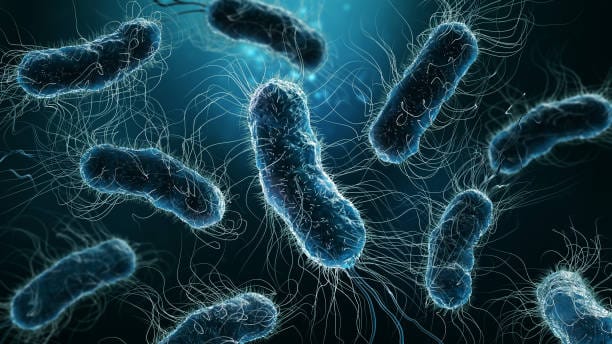
Not all bacteria are stationary. Many possess flagella—long, whip-like appendages powered by rotary motors that spin at astonishing speeds. These motors are embedded in the membrane and powered by proton flow, converting chemical energy into mechanical motion.
Pili (or fimbriae) are shorter, hair-like structures that help bacteria adhere to surfaces or to each other. Some specialized pili, known as sex pili, facilitate the transfer of DNA between cells in a process called conjugation—a microbial version of mating that spreads genetic traits like antibiotic resistance.
Endospores: Masters of Survival
In times of extreme stress, certain bacteria can form endospores—dormant, tough, and highly resistant structures that can survive for decades or even centuries. Bacillus and Clostridium species are the most famous spore-formers.
Endospores contain the bacterium’s DNA, a small amount of cytoplasm, and protective layers of protein and peptidoglycan. They can withstand radiation, desiccation, high temperatures, and chemical disinfectants. When favorable conditions return, the spore germinates and the bacterium resumes normal growth.
The Big Picture: A Simple Cell with Complex Lives
Though they lack the organelles and complexity of eukaryotic cells, bacteria are anything but primitive. They are streamlined, efficient, and highly adaptable. Their cell wall and internal structures reveal a world of biochemical ingenuity—a life reduced to its essentials but still capable of astonishing feats.
Understanding what bacteria are made of is not merely an academic exercise. It lies at the heart of medicine, agriculture, ecology, and biotechnology. It informs the design of antibiotics, the management of disease, and the harnessing of microbes for food, fuel, and industry.
These microscopic builders, scavengers, and sometimes destroyers shape our world in ways both visible and invisible. They are the originators of life’s molecular grammar and the custodians of life’s continuity. And despite their ancient lineage, they are as dynamic, diverse, and enigmatic as any creatures on Earth.
Would you like a PDF or Word version of this article for download? Or shall I help you with another microbiology topic in this same style?


