Imagine being able to watch the brain in action—not in frozen slices or after-the-fact recordings, but live, in real time, as thoughts, perceptions, and behaviors unfold. For decades, this dream has driven neuroscientists, who longed for tools powerful enough to reveal the hidden symphony of neurons firing as an animal explores, learns, and reacts to the world.
Now, researchers at the University of California, Davis, have taken a giant leap toward that dream. They have created a miniature microscope, so small and light that a mouse can carry it comfortably while moving freely. The device, called DeepInMiniscope, allows scientists to capture high-resolution, three-dimensional images of brain activity in real time—without invasive procedures that disturb natural behavior.
This breakthrough is more than just an engineering marvel; it is a window into the living brain, and with it, a doorway to new discoveries about how the mind works.
Why This Matters
The brain remains one of the greatest scientific frontiers. Despite decades of research, fundamental questions linger: How do networks of neurons create thoughts, memories, or emotions? What exactly happens in the brain when behavior shifts from curiosity to fear, or from hesitation to decision?
Studying these questions in living animals has always been difficult. Traditional microscopes and imaging devices are bulky, requiring animals to remain restrained or anesthetized. But behavior is not static—it is dynamic, flowing from moment to moment. To truly understand how brain activity shapes behavior, scientists need to see the brain while an animal is behaving naturally.
That is the promise of DeepInMiniscope: to make the invisible visible, in real time, without interrupting the delicate interplay between brain and body.
From Lensless Cameras to Brain Imaging
DeepInMiniscope did not appear overnight. It builds upon years of innovation by Weijian Yang, professor of electrical and computer engineering at UC Davis, and his team. Previously, Yang had pioneered a lensless camera system that could capture three-dimensional images from a single snapshot. This was a powerful tool for industrial applications such as robotic vision, where large objects and clean environments made image reconstruction relatively straightforward.
But biology posed a tougher challenge. Living tissue scatters light in complex ways. Neurons are small, faint, and densely packed. Extracting meaningful images from this noisy environment demanded something beyond hardware alone—it required the marriage of physics with artificial intelligence.
How DeepInMiniscope Works
The secret to DeepInMiniscope lies in its design. Instead of relying on a single large lens, the device uses a mask containing more than 100 tiny lenslets. Each lenslet captures a slightly different view of the brain. On their own, these images look incomplete. But when combined through a specially designed neural network, they reconstruct a detailed, three-dimensional picture of neuronal activity.
The computational magic comes from what Yang’s team calls an unrolled neural network. Unlike traditional deep-learning methods that often act like opaque “black boxes,” this system is designed to be interpretable and efficient. It requires only minimal training data, yet can process massive datasets at high speed.
In practice, this means that researchers can see neurons firing across large volumes of brain tissue, with fine detail, as the mouse moves naturally. It is like watching the brain’s electricity spark and flow in real time.
Small but Mighty
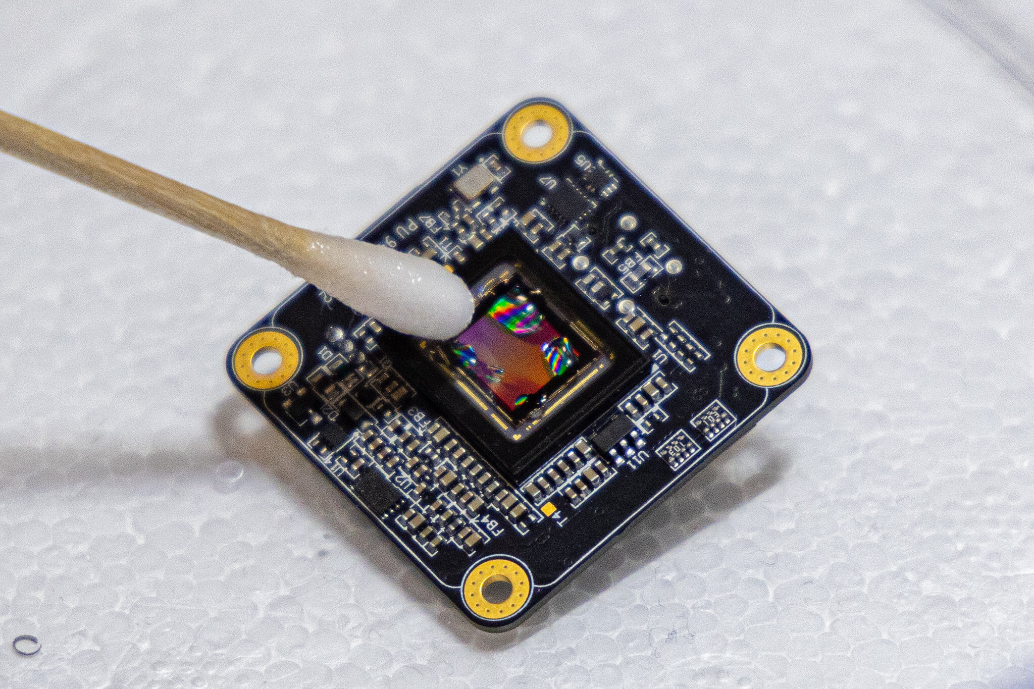
For all its power, DeepInMiniscope is remarkably compact. Measuring only about 3 square centimeters—roughly the size of a grape—and weighing just 10 grams (about the weight of four pennies), the device can be mounted on a mouse’s head without discomfort.
Earlier designs of wearable microscopes were bulky, relying on self-contained camera systems that limited miniaturization. DeepInMiniscope breaks this barrier by using an ultra-compact image sensor, essentially a bare circuit board that removes unnecessary weight and bulk.
Yang envisions an even smaller version: just 2 square centimeters, which he likens to “a tiny hat for a mouse.” His team is also working toward making the device wireless, freeing mice from the last physical tether and allowing researchers to study more natural, unconstrained behaviors.
What Scientists Can Learn
With this new tool, neuroscientists can ask questions that were once out of reach. How does a mouse’s brain process sensory information as it navigates a maze? How do neural circuits shift when the animal makes a decision, learns a new task, or responds to fear?
By correlating brain activity with real-world behaviors, researchers can begin to map how patterns of neuronal firing give rise to perception, memory, and action. These insights could extend far beyond mice. Because many fundamental principles of brain function are conserved across species, discoveries made with DeepInMiniscope may help scientists unravel the complexities of the human brain as well.
Implications for Human Health
The potential impact on medicine is profound. Many brain disorders—such as Alzheimer’s disease, epilepsy, schizophrenia, and depression—are still poorly understood at the neural-circuit level. By enabling fine-grained, real-time studies of brain activity, DeepInMiniscope could help identify the underlying mechanisms of these conditions.
Such knowledge could pave the way for new therapies and interventions—from precision-targeted drugs to advanced neurostimulation techniques. In the long run, the technology might also inspire tools for human brain imaging that are less invasive, more portable, and more accessible than today’s massive MRI and PET scanners.
The Marriage of Engineering and Neuroscience
What makes DeepInMiniscope so remarkable is that it embodies a union of disciplines. It is not just a triumph of optics or electronics. It is not just an achievement in artificial intelligence. It is the fusion of physics, engineering, computation, and biology into a single tool designed to illuminate one of the deepest mysteries in science.
As Feng Tian, a postdoctoral researcher and lead author of the paper, explained: “Our algorithm combines interpretability, efficiency, scalability and precision. It requires only a minimal amount of training data, yet it can robustly and accurately process large-scale datasets at high speed.”
This blend of technological elegance and biological relevance is what makes DeepInMiniscope more than just a gadget. It is a paradigm shift.
The Future Ahead
The journey is not over. Yang and his team continue to refine DeepInMiniscope, pushing toward smaller, lighter, and wireless versions. The vision is clear: to make brain imaging as seamless as possible, so that behavior and brain activity can be studied together in their natural, unbroken flow.
As the device improves, it may enable studies across longer timescales, capturing how the brain changes not only moment to moment, but across days, weeks, and even lifetimes. Such work could help reveal how learning reshapes neural circuits, how diseases slowly alter them, and how resilience and recovery unfold.
A Tiny Window With Immense Possibilities
At first glance, DeepInMiniscope is just a small square of electronics and optics, no larger than a grape. But in reality, it is a powerful lens through which humanity may finally begin to see the brain not as a static organ, but as a living, breathing orchestra of electrical activity.
It reminds us that the biggest breakthroughs often come in the smallest packages. And as this tiny device rides on the head of a mouse, it carries with it the hopes of neuroscientists, physicians, and patients—that one day, by understanding the brain in all its dynamic beauty, we may learn not only how it works, but how to heal it.
More information: Feng Tian et al, DeepInMiniscope: Deep-learning-powered Physics-informed Integrated Miniscope, Science Advances (2025). DOI: 10.1126/sciadv.adr6687. www.science.org/doi/10.1126/sciadv.adr6687