Urolithin A, a natural compound studied for its effects on cells and mitochondria, improved measures of diastolic heart function and reduced cardiac remodeling in a mouse model of heart failure with preserved ejection fraction. The researchers traced these effects to a specific cysteine, Cys42, on the signaling protein PKGIα, and found that urolithin A also improved the speed of contraction and relaxation in engineered human heart tissue.
The study focused on a difficult form of heart failure
Heart failure with preserved ejection fraction, or HFpEF, accounts for nearly half of all heart failure cases. The condition is marked largely by problems with diastolic function, meaning the heart has difficulty relaxing and filling properly. The problem can become more pronounced during physical activity, when the cardiovascular system needs to increase its performance.
The researchers focused on a protein called cyclic guanosine monophosphate-dependent protein kinase Iα, or PKGIα. This protein is involved in cardiovascular function and can be activated through the conventional nitric oxide and cGMP pathway. It can also be activated through other mechanisms involving oxidants.
One particular amino acid in PKGIα drew the researchers’ attention. It is cysteine 42, or Cys42. Oxidation of this cysteine can cause PKGIα to form a disulfide-linked dimer and activate signaling involved in blood pressure regulation and cardiac relaxation. Previous work had also linked Cys42-dependent PKGIα activation to phosphorylation of phospholamban, a protein involved in controlling calcium movement in heart muscle cells.
The researchers therefore tested whether compounds capable of reacting with cysteine could activate this pathway in a way that might affect HFpEF.
Several natural compounds were tested
The initial experiments examined natural compounds with potential thiol reactivity. The researchers looked at whether these compounds could promote PKGIα disulfide dimerization and increase kinase activity.
Quercetin was the compound that most clearly induced PKGIα disulfide dimerization among the compounds tested. It also increased substrate phosphorylation and worked together with cGMP to increase kinase activation. Fisetin, a structurally similar flavonoid, produced comparable PKGIα dimerization and also enhanced signaling.
Urolithin A behaved differently.
It did not produce notable PKGIα disulfide dimerization, yet it still increased substrate phosphorylation and worked together with cGMP. That combination pointed to a different mechanism of PKGIα activation.
The researchers then examined whether the compounds could relax blood vessels. Both quercetin and urolithin A produced relaxation in mouse mesenteric vessels. For urolithin A, the relaxation was reduced in vessels from mice carrying a C42S mutation in PKGIα, in which cysteine 42 is replaced by serine. Blocking soluble guanylate cyclase did not prevent the relaxation.
These experiments supported a role for Cys42 in the response to urolithin A.
Urolithin A directly modified Cys42
The researchers next examined how urolithin A could activate PKGIα without producing the usual intermolecular disulfide bond.
In a test using purified PKGIα, urolithin A directly increased kinase activity. Additional experiments showed that urolithin A was reactive toward thiols, the chemical groups found on cysteine residues.
Mass spectrometry provided more direct evidence. Urolithin A limited the disulfide dimerization of PKGIα caused by diamide and produced a detectable urolithin A adduct on Cys42.
The researchers interpreted these results as evidence that urolithin A activates PKGIα by directly modifying Cys42 rather than by producing the intermolecular disulfide bond seen with other compounds. They proposed that the modification occurs through redox cycling and formation of a thiol-reactive quinone intermediate.
This distinction was central to the study. Urolithin A was not simply reproducing the same molecular event as quercetin. Instead, it appeared to activate the same protein through a different modification of the same cysteine residue.
The modification increased signaling in heart muscle cells
The researchers then examined whether this molecular effect could influence a process directly related to cardiac relaxation.
In cardiomyocytes, urolithin A increased phosphorylation of phospholamban, or PLN. When PKGI was knocked down, the increase in PLN phosphorylation was reduced. This supported the involvement of PKGIα in the response.
PLN regulates SERCA2a, which controls the uptake of calcium into the sarcoplasmic reticulum inside heart muscle cells. The researchers noted that phosphorylation of PLN at Ser16 reduces its inhibitory effect on SERCA2a, increasing calcium uptake into the sarcoplasmic reticulum. This calcium regulation contributes to cardiac relaxation.
The findings connected the biochemical modification of Cys42 with a cellular process involved in cardiac relaxation.
The researchers next asked whether the effect would appear in an animal model of HFpEF.
Urolithin A improved diastolic function in mice
To model HFpEF, the researchers combined several stresses in mice. The animals underwent unilateral nephrectomy, received DOCA-salt treatment to promote hypertension, and were fed a high-fat diet. The researchers described this as a multihit model designed to reproduce key cardiovascular features of human HFpEF.
The experiments used both normal mice and mice carrying the C42S PKGIα mutation. After HFpEF had been established, the animals received either urolithin A or a vehicle control orally for seven days.
The treatment did not change left ventricular ejection fraction or fractional shortening. Those measures of systolic function remained preserved across the groups, and urolithin A did not produce a detectable change in heart rate.
The changes appeared instead in measures of diastolic performance.
In normal mice with HFpEF, urolithin A significantly improved global longitudinal strain, reverse longitudinal strain rate and the E/e′ ratio. The E/A ratio also improved, while the heart-rate-corrected isovolumetric relaxation time did not change.
The treatment was also associated with structural changes. Left ventricular mass and heart weight were reduced. The treated mice ran farther, while measurements of fibrosis, cardiomyocyte size and atrial natriuretic peptide expression were reduced.
The key mechanistic test came from the C42S mice.
The beneficial effects of urolithin A were absent in these mice, consistent with the requirement for PKGIα Cys42. The researchers therefore concluded that urolithin A limited cardiac dysfunction and remodeling in this HFpEF model through C42-dependent PKGIα activation.
The human heart tissue experiments showed faster contraction and relaxation
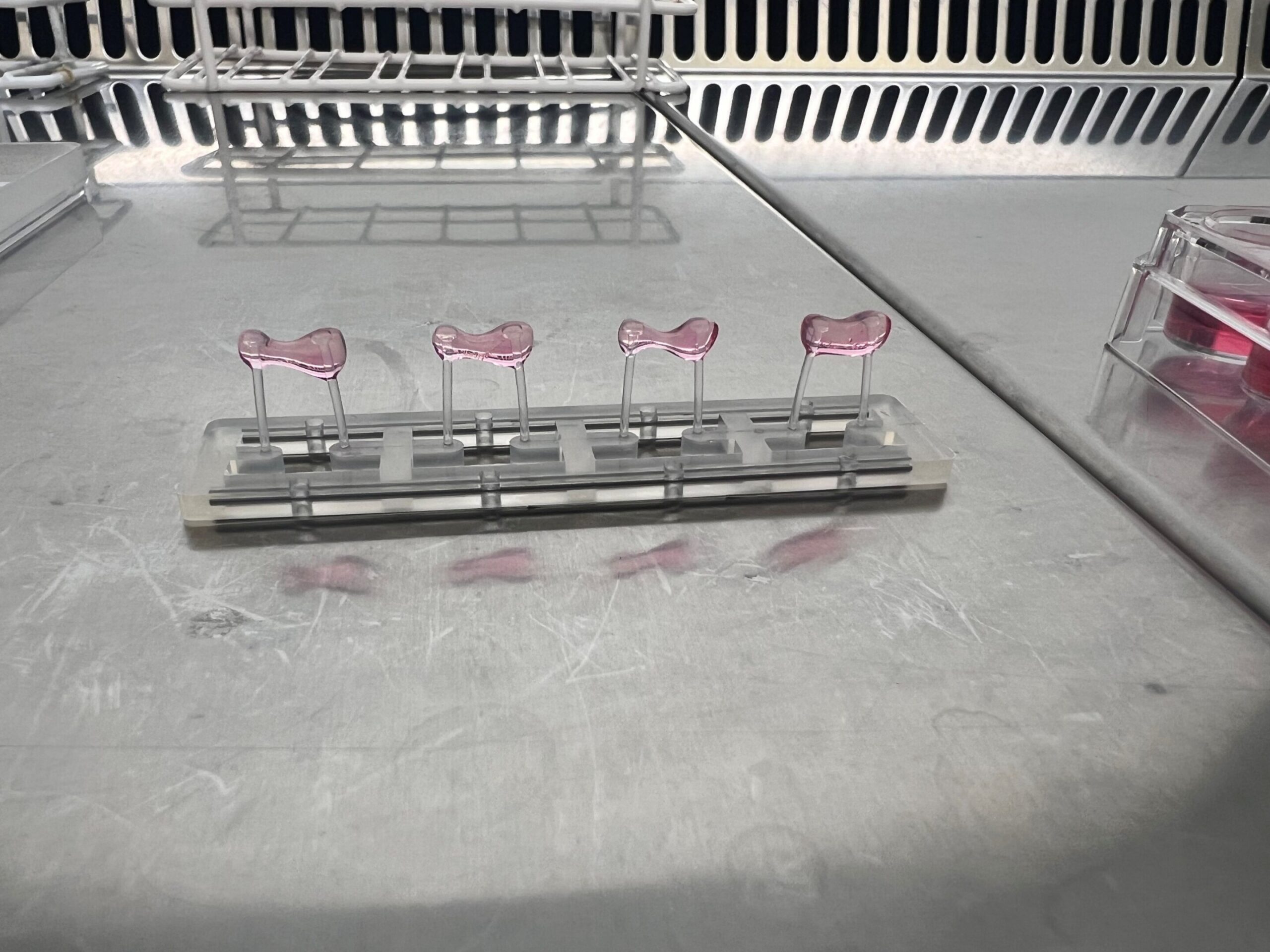
The researchers also tested urolithin A in engineered human heart tissue. The tissue was made from cardiomyocytes generated from a single human induced pluripotent stem cell line.
Urolithin A did not change maximum force generation, and no arrhythmias were detected in the experiments. But it did change the timing of both contraction and relaxation.
After 120 minutes of treatment, measures of relaxation time decreased. T20% fell by 32%, T50% by 36% and T90% by 40%. Measures of contraction time also decreased, with T20% falling by 49%, T50% by 48% and T90% by 34%.
The researchers said these results were consistent with improved calcium handling, likely through PKGI-dependent phosphorylation of phospholamban. They concluded that urolithin A enhanced contraction and relaxation parameters in the engineered human cardiac tissue.
The findings point to Cys42 as the key target
The experiments brought together several levels of evidence.
Urolithin A directly activated PKGIα in biochemical assays. It reacted with thiols and produced an identifiable modification of Cys42. It increased PLN phosphorylation in cardiomyocytes in a PKGI-dependent manner. It relaxed blood vessels in a manner that depended on Cys42. And in mice with HFpEF, its effects on cardiac function and remodeling depended on the same cysteine.
The mouse model was designed to combine hypertension and metabolic stress. It produced preserved ejection fraction together with reduced global longitudinal strain, impaired reverse longitudinal strain rate, reduced exercise capacity and increased interstitial fibrosis. The researchers reported that this phenotype was also reproduced in an independent facility and on a different mouse background in earlier work.
The researchers also noted an important limitation of the animal work. The experiments focused on male mice, even though HFpEF disproportionately affects women. They said whether this model accurately reproduces the HFpEF phenotype in females remains to be determined.
The authors also cautioned that transmitral Doppler measurements in mice need careful interpretation because mice have extremely high heart rates and compressed diastolic intervals compared with humans. They considered deformation-based measurements more informative for mechanistic analysis in this setting.
The study does not establish a treatment for people with HFpEF
The human experiments in this study used engineered heart tissue rather than patients. The main therapeutic testing was performed in the mouse HFpEF model, while the human-derived tissue experiments examined contraction and relaxation kinetics.
The researchers pointed to previous human studies reporting that orally administered urolithin A is bioavailable and has a favorable safety profile. They also noted that earlier human work had examined its effects on mitochondrial and cellular health and on muscle endurance. Those findings provided part of the rationale for examining urolithin A in the HFpEF model.
But the present study’s direct evidence for HFpEF came from the preclinical model and engineered human heart tissue.
The researchers therefore focused their conclusion on the molecular target they identified. They described Cys42 in PKGIα as a potential therapeutic target and urolithin A as a previously unidentified activator of this protective mechanism. They also proposed that more potent and selective compounds capable of targeting the same cysteine could be developed as another way of pursuing this mechanism.
The study was published in Science Advances.