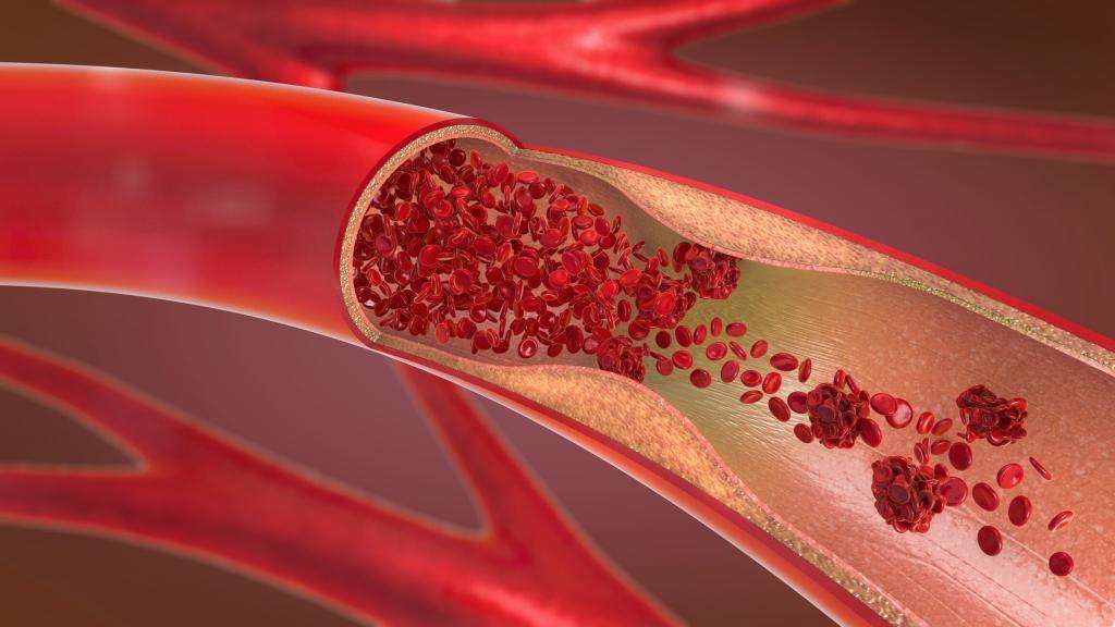
Every moment of your life, blood is traveling through your body. It carries oxygen from your lungs to tissues that need it, delivers nutrients absorbed from food, transports hormones and other chemical signals, carries carbon dioxide and metabolic waste away, helps regulate body temperature, and participates in the immune and clotting systems that protect you from injury and infection.
What makes this possible is the circulatory system, also called the cardiovascular system. It is a vast, branching network made up primarily of the heart, blood, and blood vessels. Far from being a simple set of pipes with a pump attached, it is a dynamic system that constantly adjusts blood flow to match the changing needs of different tissues.
Your heart may beat faster during exercise, while blood vessels in working muscles widen to receive more blood. At the same time, vessels supplying less immediately active tissues can constrict. When you stand up, specialized sensors help your circulation respond to gravity. When you become overheated, blood flow near the skin increases to help release heat. When you are injured, a carefully coordinated clotting response helps prevent excessive blood loss.
The remarkable feature of this system is not simply that blood moves. It is that blood moves in the right direction, at the right pressure, through the right tissues, and in quantities that continually change according to what the body requires.
What the circulatory system actually does
The circulatory system connects virtually every part of the body. Its most familiar job is transportation, but its responsibilities extend much further.
Red blood cells carry oxygen from the lungs to tissues and return with some of the carbon dioxide produced by cellular metabolism. Blood plasma carries nutrients such as glucose, amino acids, electrolytes, and many other dissolved substances. It also transports hormones released by endocrine organs to distant tissues where those signals have their effects.
Blood also serves as a waste-transport system. Carbon dioxide travels to the lungs to be exhaled, while substances destined for removal by the kidneys, liver, or other organs are carried through the bloodstream to those locations.
The circulatory system helps maintain the body’s internal environment, a process known as homeostasis. By distributing heat, water, electrolytes, hormones, and other substances, it helps keep conditions within cells and tissues within ranges compatible with life.
It also provides defense. White blood cells circulate through the blood and can enter tissues when needed. Proteins and other components of the immune system travel through the circulation as well. Meanwhile, platelets and clotting proteins help stop bleeding when a blood vessel is damaged.
These functions depend on constant movement. A cell cannot receive oxygen or nutrients merely because they exist somewhere in the body. They must be transported close enough for exchange to occur.
The heart is the system’s muscular engine
At the center of the circulatory system is the heart, a muscular organ located in the chest between the lungs. Its job is to generate the pressure that drives blood through the vessels.
The human heart has four chambers. The two upper chambers, called the atria, receive blood. The two lower chambers, called the ventricles, pump blood out of the heart.
The right side of the heart handles blood returning from the body. This blood has relatively little oxygen because body tissues have extracted oxygen and released carbon dioxide. The right ventricle pumps this blood to the lungs, where carbon dioxide is released and oxygen enters the bloodstream.
The oxygen-rich blood then returns to the left side of the heart. The left ventricle pumps it into the aorta, the body’s largest artery, beginning its journey through the systemic circulation to tissues throughout the body.
This arrangement creates two linked circuits. The pulmonary circulation carries blood between the heart and lungs. The systemic circulation carries blood between the heart and the rest of the body.
The heart therefore performs two related pumping jobs. The right ventricle sends blood through the lungs, while the left ventricle sends blood through the much larger systemic circuit.
How one heartbeat moves blood
A heartbeat is not simply the heart squeezing once. It is a coordinated sequence of electrical and mechanical events.
The heart’s normal rhythm begins with electrical activity generated by specialized cells in the sinoatrial node, often called the heart’s natural pacemaker. This electrical signal spreads through the atria, causing them to contract and push blood into the ventricles.
The signal then passes through the atrioventricular node and travels through specialized conduction pathways into the ventricles. This causes the ventricles to contract.
When the ventricles contract, pressure inside them rises. Blood is pushed through the appropriate valves and into the pulmonary artery or aorta. When the ventricles relax, their pressure falls and they fill again.
Heart valves are crucial because they help keep blood moving in one direction. The tricuspid and mitral valves separate the atria from the ventricles, while the pulmonary and aortic valves control the exits from the ventricles.
The familiar sounds of the heartbeat are largely associated with the closing of these valves. The first heart sound occurs when the valves between the atria and ventricles close as the ventricles begin contracting. The second occurs when the pulmonary and aortic valves close as the ventricles begin relaxing.
This cycle repeats continuously. At rest, a typical adult heart beats roughly 60 to 100 times per minute, although normal heart rates vary with circumstances, fitness, age, medications, and other factors.
The electrical system keeps the pumping coordinated
The heart could not pump efficiently if its muscle cells contracted randomly. Its electrical conduction system provides the timing that coordinates the chambers.
The sinoatrial node normally initiates each heartbeat. The signal spreads across the atria and reaches the atrioventricular node, where conduction briefly slows. That delay gives the ventricles time to fill before they contract.
From there, the electrical impulse travels through the bundle of His, its branches, and Purkinje fibers, distributing the signal throughout the ventricular muscle.
This electrical organization allows the heart to behave as a coordinated pump rather than as a collection of independent muscle cells.
The nervous system also influences the heart. The sympathetic nervous system can increase heart rate and the strength of contraction during situations such as exercise or stress. The parasympathetic nervous system, particularly through the vagus nerve, generally slows the heart.
Importantly, the heart does not need the brain to send an individual command for every beat. Its own specialized electrical cells generate the basic rhythm, while the nervous and hormonal systems adjust that rhythm according to the body’s circumstances.
Arteries, veins, and capillaries have different jobs
Blood vessels are not interchangeable tubes. Their structures reflect the different pressures and functions they encounter.
Arteries carry blood away from the heart. The largest artery is the aorta, which branches repeatedly into progressively smaller arteries and then arterioles. With every heartbeat, the left ventricle sends a surge of blood into the aorta, creating pressure within the arterial system.
Arterial walls contain substantial amounts of smooth muscle and elastic tissue. The larger arteries can stretch when blood is ejected from the heart and recoil afterward. This helps smooth out the pulsatile output of the heart and maintain blood flow between individual heartbeats.
Arterioles are smaller vessels with a particularly important role in controlling blood distribution. Their muscular walls can constrict or relax, changing their diameter and therefore influencing how much blood reaches downstream tissues.
Capillaries are the smallest blood vessels. Their walls are extremely thin, generally only one cell layer thick. This makes them well suited for exchanging oxygen, carbon dioxide, nutrients, water, hormones, and waste products between blood and surrounding tissues.
After passing through capillaries, blood enters venules and then veins. Veins return blood to the heart.
Because blood pressure is much lower in the venous system than in the arteries, veins have thinner walls. Many veins, particularly in the limbs, contain valves that help prevent blood from flowing backward.
Why capillaries are so important
The body’s cells do not normally exchange most substances directly with large arteries or veins. The critical interface is the capillary network.
A single tissue may contain an extensive network of microscopic capillaries. Their enormous combined surface area provides opportunities for exchange between blood and the fluid surrounding cells.
Oxygen moves from blood into tissues when conditions favor that movement, while carbon dioxide moves in the opposite direction. Glucose and other nutrients can leave the blood and enter tissues. Metabolic wastes can enter the bloodstream for transport elsewhere.
Not every capillary is equally active at every moment. Local mechanisms can alter blood flow through different vascular networks depending on tissue demands.
During exercise, for example, working skeletal muscles require substantially more oxygen and nutrients and produce more metabolic waste and heat. Blood flow to those muscles can increase dramatically through changes in local blood vessels.
The circulation therefore does not simply distribute the same amount of blood everywhere. It continuously reallocates blood according to physiological needs.
Blood pressure is the force that drives circulation
Blood moves because there are differences in pressure within the circulatory system. The heart creates much of that pressure by contracting, while the properties of the blood vessels influence how pressure and flow are maintained.
Blood pressure is usually discussed in terms of two numbers. Systolic pressure is the higher pressure produced during ventricular contraction. Diastolic pressure is the lower pressure during ventricular relaxation.
Blood pressure is not the same throughout the circulatory system. It is highest in the large arteries and progressively falls as blood travels through smaller vessels and eventually returns to the heart through the veins.
This pressure difference is essential. Blood flows from regions of higher pressure toward regions of lower pressure.
The relationship between pressure and flow is more complicated than simply saying that higher pressure always means more blood flow. Vessel diameter and resistance are extremely important. Small changes in the radius of a blood vessel can produce large changes in the resistance to flow.
This is one reason arterioles are such powerful regulators of circulation. By changing their diameter, they can substantially alter resistance and influence both local blood flow and overall blood pressure.
What determines how much blood the heart pumps?
The amount of blood pumped by the heart each minute is called cardiac output.
Cardiac output depends mainly on two factors: heart rate and stroke volume. Heart rate is the number of beats per minute. Stroke volume is the amount of blood ejected by a ventricle with each beat.
For example, a heart beating 70 times per minute and ejecting 70 milliliters per beat would have a cardiac output of about 4.9 liters per minute.
At rest, an adult’s cardiac output is often around five liters per minute, although the actual value varies considerably. During strenuous exercise, cardiac output can increase several-fold as the heart beats faster and ejects more blood with each contraction.
Stroke volume is influenced by how much blood fills the ventricle, how strongly the heart muscle contracts, and the resistance against which the ventricle must pump.
The relationship between filling and contraction is especially important. Within normal physiological limits, when more blood returns to the heart and stretches the ventricular muscle, the heart generally contracts more forcefully and ejects more blood. This is known as the Frank-Starling mechanism and helps the heart automatically adjust its output to the amount of blood returning to it.
How blood gets back to the heart
Moving blood out of the heart is only half the problem. The circulation must also return blood efficiently, particularly from the feet and legs, where gravity works against the upward journey.
Veins act as important reservoirs because they can hold a substantial proportion of the body’s blood volume. But venous return is assisted by several mechanisms.
When skeletal muscles contract, particularly during walking, they can compress nearby veins and help push blood toward the heart. Valves inside many veins help keep that blood moving in the correct direction.
Breathing also contributes. Changes in pressure within the chest during normal respiration can help draw venous blood toward the heart.
These mechanisms are especially important when a person is standing. Without effective regulation, gravity would cause blood to pool in the lower body, reducing the amount returning to the heart and potentially lowering blood flow to the brain.
The body constantly adjusts blood flow
The circulatory system is remarkably responsive. It must cope with changes in posture, temperature, physical activity, emotional state, digestion, sleep, illness, and many other conditions.
Blood vessels can constrict or dilate in response to signals from the nervous system, hormones, and local chemical conditions.
When a person exercises, the cardiovascular system responds in several coordinated ways. Heart rate rises, the heart contracts more strongly, and blood flow increases to active muscles. Blood vessels within working muscles can dilate, while other vascular beds may receive a smaller share of the available blood flow.
At the same time, the body has to manage heat. Active muscles generate heat, and increased blood flow to the skin can help transfer that heat to the environment. In hot conditions, this mechanism becomes particularly important.
During digestion, blood flow to the gastrointestinal organs changes as those tissues become more active. During sleep, cardiovascular demands change again.
This constant redistribution illustrates a central principle of physiology: circulation is regulated according to function rather than simply divided equally among organs.
How the body knows when blood pressure changes
The body has specialized pressure sensors called baroreceptors, located primarily in the walls of major arteries in the neck and chest.
These receptors detect stretching of the arterial walls, which provides information about blood pressure. Signals from them reach cardiovascular control centers in the brainstem.
If blood pressure suddenly falls, as can happen when a person stands up quickly, the body can respond by increasing sympathetic nervous activity. The heart may beat faster and more forcefully, while certain blood vessels constrict. These changes help restore blood pressure and maintain blood flow to vital organs.
If blood pressure rises, the opposite pattern can occur.
This feedback system operates continuously and rapidly. It is one reason healthy circulation can accommodate ordinary changes in body position without causing persistent disruption of blood flow to the brain.
Blood itself is more than a red liquid
Blood is a specialized connective tissue consisting of cells suspended in a fluid called plasma.
Plasma is mostly water, but it also contains proteins, electrolytes, nutrients, hormones, gases, waste products, and many other substances.
Red blood cells are the most abundant blood cells. Their main function is to transport oxygen using hemoglobin, an iron-containing protein. Hemoglobin also contributes to the transport of carbon dioxide and helps buffer acids in the blood.
White blood cells are involved in immune defense. Different types have different roles, ranging from recognizing and destroying infectious organisms to coordinating immune responses.
Platelets are small cell fragments that are essential to blood clotting. When a blood vessel is damaged, platelets become activated and participate in forming a temporary plug. A series of clotting reactions then helps reinforce the developing clot with a protein mesh called fibrin.
Clotting must be carefully regulated. Too little clotting can allow dangerous bleeding, while excessive or inappropriate clot formation can obstruct blood vessels.
Why blood is red
The color of blood comes primarily from hemoglobin inside red blood cells.
Oxygenated blood is typically bright red because oxygen binding changes the way hemoglobin absorbs and reflects light. Blood with less oxygen is darker red.
Blood in veins is therefore dark red rather than blue. The bluish appearance sometimes associated with veins beneath the skin results from the way light interacts with skin and underlying tissues, not because venous blood is blue.
The circulation is a closed loop
It is tempting to imagine blood as traveling through a single route from the heart to the body and then directly back again. In reality, the circulation branches extensively.
Blood leaving the left ventricle travels through the aorta and progressively smaller arteries, arterioles, and capillaries before returning through venules and veins to the right side of the heart.
From there, it travels through the pulmonary arteries to the lungs, passes through pulmonary capillaries, and returns through the pulmonary veins to the left atrium.
The pulmonary arteries are an important reminder that the terms artery and vein describe direction rather than oxygen content. An artery carries blood away from the heart, regardless of whether that blood is oxygen-rich or oxygen-poor. A vein carries blood toward the heart.
Thus, pulmonary arteries carry relatively oxygen-poor blood, while pulmonary veins carry oxygen-rich blood.
The lungs complete the blood’s oxygen journey
The heart and lungs work as a tightly integrated unit.
When oxygen-poor blood reaches the pulmonary capillaries, it flows alongside air-filled spaces in the lungs. Oxygen moves across the thin respiratory membrane and enters the blood. Carbon dioxide moves from the blood into the air spaces and is then exhaled.
This exchange works because of differences in the concentrations and partial pressures of the gases on the two sides of the membrane.
Once oxygen has entered the bloodstream, much of it binds to hemoglobin. This allows the blood to transport far more oxygen than could be carried simply dissolved in plasma.
The oxygenated blood returns to the left side of the heart, which then distributes it throughout the body.
Why the circulation needs four chambers
The four-chambered design of the human heart keeps pulmonary and systemic circulation connected while preventing the two flows from mixing under normal conditions.
The right side pumps blood through the lungs. The left side pumps blood through the rest of the body.
The left ventricle has a thicker muscular wall than the right ventricle because it must generate substantially higher pressure to move blood through the systemic circulation.
The right ventricle pumps into the pulmonary circulation, which is a shorter and lower-resistance circuit. Its walls therefore do not need to generate the same pressure.
This division allows the lungs to receive blood at pressures appropriate for efficient gas exchange while the systemic circulation receives the higher pressure needed to deliver blood throughout the body.
The coronary circulation keeps the heart itself alive
The heart pumps blood throughout the body, but the blood inside its chambers does not directly provide most of the oxygen needed by the heart muscle itself.
Instead, the myocardium receives blood through the coronary arteries, which branch from the beginning of the aorta.
The heart muscle is highly metabolically active and requires a continuous oxygen supply. Coronary blood flow therefore plays a critical role in maintaining normal cardiac function.
If a coronary artery becomes severely narrowed or blocked, the region of heart muscle supplied by that vessel may not receive enough oxygen. Temporary oxygen shortage can cause ischemia and may produce chest discomfort or other symptoms. Prolonged interruption of blood supply can cause myocardial infarction, commonly called a heart attack, in which heart muscle cells are injured or die.
The brain has its own demanding blood supply
The brain represents only a small fraction of total body mass but consumes a substantial amount of energy. It depends heavily on a continuous supply of oxygen and glucose.
The brain receives blood through major arteries that enter the skull and branch into an intricate vascular network. Specialized mechanisms help regulate cerebral blood flow so that the brain receives an appropriate supply despite changes in overall blood pressure.
Because brain cells are highly dependent on continuous oxygen and nutrient delivery, interruption of cerebral blood flow can cause rapid injury. A stroke can occur when blood flow to part of the brain is blocked, or when a blood vessel in the brain ruptures and causes bleeding.
The consequences depend on which part of the brain is affected and how extensive the injury is.
The kidneys use circulation to regulate the body’s chemistry
The kidneys receive a remarkably large blood supply relative to their size. This circulation allows them to filter plasma and regulate the composition of the internal environment.
Within the kidneys, specialized capillary networks participate in filtration. The resulting fluid is processed through microscopic tubules, where useful substances and water can be reabsorbed while other substances are excreted.
The kidneys also play an important role in blood pressure regulation. They influence the body’s sodium and water balance and participate in hormonal systems that regulate vascular tone and blood volume.
This creates a two-way relationship: circulation supplies the kidneys with the blood they need to perform their functions, while the kidneys help regulate the very variables that influence circulation.
The liver receives blood from two major sources
The liver has a distinctive circulatory arrangement. It receives oxygenated blood from the hepatic artery and nutrient-rich blood from the hepatic portal vein.
The portal vein carries blood from much of the digestive tract to the liver. Nutrients absorbed from food therefore pass through the liver before entering the general systemic circulation.
This arrangement allows the liver to process, store, transform, or detoxify many substances absorbed from the digestive system.
The liver also produces important blood proteins, including many proteins involved in blood clotting, and plays major roles in metabolism and the handling of numerous substances carried in the bloodstream.
What happens when a blood vessel is damaged?
The body has a sophisticated response to vascular injury.
When the inner lining of a blood vessel is disrupted, platelets can adhere to the damaged area and become activated. They release and respond to chemical signals that recruit additional platelets.
At the same time, proteins circulating in the blood activate a series of reactions known collectively as the coagulation cascade. These reactions ultimately generate fibrin, which forms a mesh that stabilizes the platelet plug.
The damaged vessel can also constrict, reducing blood loss.
Once repair is sufficiently advanced, the body has mechanisms for breaking down and removing the clot. The ability to form a clot when needed and dismantle it afterward is an important part of maintaining vascular health.
Why arteries develop disease
Blood vessels are living tissues rather than inert pipes. Their inner lining, called the endothelium, participates actively in regulating blood flow, inflammation, clotting, and interactions between the blood and vessel wall.
Atherosclerosis is a disease process in which fatty deposits, inflammatory cells, connective tissue, and other materials accumulate within the walls of arteries. Over time, these changes can form plaques that narrow arteries or alter their structure.
A plaque can sometimes become unstable and rupture. This can trigger clot formation on its surface and suddenly obstruct blood flow.
Atherosclerosis can affect arteries supplying the heart, brain, legs, and other organs. Its consequences therefore depend partly on which vascular territories are affected.
High blood pressure can also damage blood vessels over time. Persistent pressure places mechanical stress on arterial walls and contributes to changes that can make vessels less healthy and increase the risk of cardiovascular complications.
Why veins can become enlarged
Veins have lower internal pressure than arteries and are designed to return blood to the heart. When the walls or valves of certain veins become less effective, blood can pool within them.
Varicose veins are enlarged, twisted veins that most commonly occur in the legs. Standing for long periods, inherited factors, aging, pregnancy, and other influences can contribute to their development.
Not every visible vein represents disease. Veins near the skin can become more prominent because of genetics, body composition, temperature, physical activity, or other normal variations.
The broader physiological issue is venous return: blood must overcome gravity and travel back toward the heart efficiently.
The circulation also helps control body temperature
Blood is an important heat-distribution system.
Metabolic reactions continuously generate heat, especially in active muscles and organs. Blood carries heat away from warmer tissues and distributes it throughout the body.
When the body needs to lose heat, blood vessels near the skin can widen. More warm blood then travels close to the body’s surface, allowing heat to transfer to the surrounding environment.
When conserving heat is more important, skin blood vessels can constrict, reducing heat transfer from the body’s core to the environment.
This is one reason skin color and temperature can change with environmental conditions, exercise, and emotional responses. The changes often reflect alterations in blood flow near the skin.
Exercise puts the circulation to work
Physical activity provides a clear demonstration of cardiovascular regulation.
As exercise begins, muscles require more ATP, the cellular energy currency. Producing that energy aerobically requires increased oxygen delivery and generates additional carbon dioxide and heat.
The cardiovascular system responds by increasing cardiac output. The heart beats faster and generally pumps more blood with each beat. Blood vessels supplying active muscles adjust to accommodate increased flow.
Meanwhile, breathing becomes faster and deeper, increasing the delivery of oxygen to the lungs and removal of carbon dioxide.
Regular physical activity can produce long-term adaptations in the cardiovascular system. In many physically trained people, the heart becomes more efficient at pumping blood, and the body becomes better able to deliver and use oxygen during exercise.
Why the pulse can be felt
When the left ventricle ejects blood into the aorta, it creates a pressure wave that travels through the arterial system.
That wave can be detected where an artery lies close enough to the skin to be compressed against underlying tissue. Common locations include the wrist and side of the neck.
The pulse therefore reflects the rhythmic activity of the heart as transmitted through the arteries. It is not simply the physical movement of the same drop of blood all the way from the heart to the point where you feel it.
The pulse can provide useful information about heart rate and rhythm, although assessing cardiovascular health requires more than feeling the pulse alone.
What makes the system so efficient?
Several features work together to make circulation effective.
The heart generates pressure. The arteries distribute blood under relatively high pressure. Arterioles regulate resistance and local flow. Capillaries provide enormous surfaces for exchange. Veins return blood at lower pressure and use valves, muscle contractions, and respiratory movements to assist that return.
The branching architecture of the vascular network is equally important. One large artery can divide into smaller arteries, then arterioles, and ultimately countless capillaries. This arrangement allows the circulation to reach tissues throughout the body while keeping individual capillary distances short.
The system also has extraordinary regulatory flexibility. Blood flow can be redirected within seconds or minutes as circumstances change.
Perhaps most importantly, circulation is integrated with virtually every other major physiological system. The lungs load blood with oxygen. The digestive system supplies nutrients. The kidneys regulate fluid and electrolyte balance. The liver processes substances carried in the blood. The nervous and endocrine systems alter cardiovascular function. Muscles and other tissues change their blood supply according to metabolic demand.
What happens when circulation stops?
The dependence of the body on circulation becomes especially clear when blood flow is interrupted.
If the heart stops pumping effectively, blood pressure falls rapidly and organs stop receiving adequate oxygen and nutrients. The brain is particularly vulnerable because its energy requirements are high and it has limited capacity to store the substrates needed for continuous metabolism.
Likewise, if an artery supplying a particular tissue becomes blocked, that tissue can become ischemic because its cells are no longer receiving enough blood.
The severity depends on the location and extent of the interruption, how long it lasts, and whether alternative pathways can supply some blood.
The body does have collateral vessels and other adaptations that can sometimes provide alternate routes around a narrowed or blocked vessel. But these routes vary substantially among individuals and tissues and cannot always compensate for a major obstruction.
The circulation changes throughout life
The cardiovascular system develops continuously from early embryonic life onward. The fetal circulation has specialized features because a fetus does not breathe air with its lungs. Oxygen and nutrients are supplied through the placenta, and fetal blood follows a circulation adapted to that environment.
At birth, breathing begins and the circulation undergoes major changes. Blood flow through the lungs increases, and temporary fetal pathways normally close or become functionally insignificant.
During childhood and adolescence, the heart and blood vessels grow along with the body. In adulthood, cardiovascular structure and function continue to respond to activity, hormones, aging, and disease.
With aging, blood vessels commonly become less elastic, and the heart and vascular system may become less able to respond to certain stresses. These changes are influenced not only by age itself but also by lifelong exposures, genetics, physical activity, diet, smoking, blood pressure, metabolic health, and other factors.
Keeping the circulation healthy
Because the cardiovascular system is interconnected with the rest of the body, its long-term health is influenced by many aspects of everyday life.
Regular physical activity supports cardiovascular fitness and helps the body use its circulation efficiently. A nutritious eating pattern, adequate sleep, avoiding tobacco, maintaining a healthy body weight when possible, and managing conditions such as high blood pressure, diabetes, and abnormal cholesterol can all affect cardiovascular risk.
Some cardiovascular problems develop without obvious symptoms for years. High blood pressure, for example, may cause no noticeable symptoms even while gradually increasing the strain on blood vessels and the heart.
That is why routine health care and appropriate screening can matter even when a person feels well.
Symptoms such as persistent or severe chest pressure, unexplained shortness of breath, sudden weakness or numbness, difficulty speaking, fainting, or other abrupt neurological changes can signal medical emergencies and warrant immediate medical attention.
The remarkable part of circulation is not just the heart’s ability to beat thousands of times each day. It is the coordination of billions of cells, millions of vessels, chemical signals, electrical impulses, pressure changes, and feedback mechanisms that keep blood moving continuously while adapting to the body’s changing needs.
From the largest artery to a microscopic capillary, every part of the network contributes to the same fundamental task: bringing the body’s tissues what they need and carrying away what they no longer need.