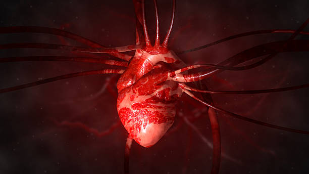
The human heart is the central powerhouse of life, an extraordinary muscular organ that beats tirelessly from before birth until the final moment of existence. It drives the continuous circulation of blood throughout the body, delivering oxygen and nutrients to every cell and carrying away carbon dioxide and metabolic waste. Without the heart’s rhythmic pumping, the body’s organs and tissues would quickly perish, for it is through this ceaseless flow of blood that life is sustained.
More than just a mechanical pump, the human heart is a masterpiece of biological engineering—a marvel of precision, resilience, and coordination. It operates in harmony with the lungs, blood vessels, and the nervous system to maintain homeostasis. Its beat reflects both the body’s physical demands and the subtle influences of emotion, stress, and environment. Every heartbeat tells a story of adaptation, survival, and the delicate balance that sustains life on Earth.
To understand the heart is to understand the essence of human physiology. Its function lies at the crossroads of biology, chemistry, and physics, combining electrical impulses, muscular contraction, and fluid dynamics into a symphony that repeats over 100,000 times each day.
The Structure of the Human Heart
The human heart is a four-chambered organ roughly the size of a clenched fist, weighing about 250 to 350 grams in an average adult. It is located slightly to the left of the midline in the thoracic cavity, nestled between the lungs and protected by the ribcage. The heart sits in a fluid-filled sac called the pericardium, which cushions and lubricates it during contraction and relaxation.
Anatomically, the heart is divided into two sides: the right and the left. Each side functions as a separate pump, serving different circuits of circulation. The right side pumps deoxygenated blood to the lungs for oxygenation—a process known as pulmonary circulation. The left side pumps oxygen-rich blood to the rest of the body through systemic circulation.
Each side contains two chambers: an atrium and a ventricle. The atria are the upper chambers that receive incoming blood, while the ventricles are the lower chambers responsible for pumping blood out of the heart. The right atrium collects deoxygenated blood from the body through the superior and inferior vena cava, passing it into the right ventricle, which sends it to the lungs via the pulmonary arteries. After oxygenation, the blood returns through the pulmonary veins into the left atrium, and then into the left ventricle—the most powerful chamber—which propels it through the aorta to every organ and tissue.
Between these chambers and major vessels lie four crucial valves: the tricuspid, pulmonary, mitral, and aortic valves. These ensure that blood flows in one direction only, preventing any backward leakage. The opening and closing of these valves produce the familiar “lub-dub” sounds heard through a stethoscope.
The Microscopic Anatomy of the Heart
At the microscopic level, the heart is composed of specialized muscle tissue known as cardiac muscle or myocardium. Unlike skeletal muscle, which can rest, cardiac muscle must contract continuously throughout life. Its cells, called cardiomyocytes, are uniquely adapted for endurance and synchronization. They contain abundant mitochondria to supply the immense energy required for constant activity, and they are interconnected by intercalated discs that allow electrical impulses to spread rapidly, ensuring coordinated contractions.
The heart also possesses its own electrical conduction system, a network of pacemaker cells that generate and transmit impulses without external stimulation. This autonomy allows the heart to beat independently of the brain, though it remains influenced by the nervous system and hormones.
The Electrical System of the Heart
The rhythmic contraction of the heart is governed by its intrinsic conduction system—a self-regulating electrical network that initiates and coordinates each heartbeat. The process begins in the sinoatrial (SA) node, a cluster of specialized cells located in the right atrium. Known as the heart’s natural pacemaker, the SA node generates electrical impulses at a rate of 60 to 100 times per minute in a healthy adult.
These impulses spread across the atria, causing them to contract and push blood into the ventricles. The signal then reaches the atrioventricular (AV) node, which acts as a gateway, briefly delaying transmission to ensure that the ventricles fill completely before contracting. From the AV node, the impulse travels along the bundle of His, which divides into right and left bundle branches that extend into the ventricles. Finally, the signal disperses through Purkinje fibers, triggering the synchronized contraction of ventricular muscle and the ejection of blood.
This electrical activity can be measured and recorded through an electrocardiogram (ECG or EKG), which provides valuable information about the heart’s rhythm, rate, and overall health. Disturbances in this electrical pattern can lead to arrhythmias—irregular heartbeats that can be benign or life-threatening, depending on their nature.
The Cardiac Cycle: The Rhythm of Life
Every heartbeat represents one complete cardiac cycle, consisting of two main phases: systole (contraction) and diastole (relaxation). During systole, the ventricles contract, propelling blood into the pulmonary artery and the aorta. During diastole, the ventricles relax and fill with blood from the atria. This continuous alternation between contraction and relaxation ensures an unbroken flow of blood throughout the body.
The cardiac cycle is regulated not only by the heart’s intrinsic pacemaker but also by neural and hormonal influences. The autonomic nervous system plays a vital role: the sympathetic branch accelerates heart rate during stress or exertion, while the parasympathetic branch slows it down during rest. Hormones such as adrenaline, thyroid hormones, and cortisol can also modify cardiac function according to the body’s needs.
The heart rate and stroke volume (the amount of blood pumped per beat) determine cardiac output—the total volume of blood the heart ejects per minute. In an average adult at rest, cardiac output is about five liters per minute, matching the total blood volume of the body. During intense exercise, this can increase fivefold or more to meet the heightened demand for oxygen and nutrients.
Circulatory Pathways: The Heart and Its Vessels
The heart does not work in isolation; it is the central hub of an extensive vascular network. Arteries carry blood away from the heart, veins return it, and capillaries connect the two systems at the tissue level.
In pulmonary circulation, the right ventricle pumps deoxygenated blood through the pulmonary arteries to the lungs. There, carbon dioxide is released and oxygen is absorbed into the bloodstream. The oxygenated blood then returns via pulmonary veins to the left atrium.
In systemic circulation, the left ventricle sends oxygen-rich blood through the aorta and branching arteries to every organ. Capillaries deliver oxygen and nutrients while collecting waste products. Deoxygenated blood then flows into veins, converging into the superior and inferior vena cava, which empty back into the right atrium—completing the circuit.
This dual circulation ensures that oxygen exchange and nutrient delivery occur efficiently. The heart and vessels also adjust dynamically to maintain blood pressure and flow according to activity level, posture, and environmental conditions.
The Heart’s Energy Demands and Metabolism
The heart is one of the most energy-demanding organs in the body. Despite weighing less than half a kilogram, it consumes about 5% of the body’s oxygen supply even at rest. Cardiac muscle relies primarily on aerobic metabolism, meaning it requires a constant supply of oxygen and nutrients—mainly fatty acids and glucose—to produce adenosine triphosphate (ATP), the cellular energy currency.
The coronary arteries, which branch off from the base of the aorta, supply this oxygen-rich blood to the heart muscle itself. If these arteries become narrowed or blocked, the resulting oxygen deprivation can cause ischemia or even a heart attack (myocardial infarction). Because the heart cannot tolerate long periods without oxygen, uninterrupted coronary blood flow is vital for survival.
The Heart in the Context of the Human Body
The heart’s function cannot be fully understood without considering its integration into the broader physiological system. It works in close partnership with the lungs, kidneys, and brain to regulate oxygen delivery, fluid balance, and blood pressure. The respiratory system ensures a continuous exchange of gases, the kidneys control blood volume and electrolyte composition, and the brain—especially through the medulla oblongata—monitors and adjusts cardiac performance through neural feedback.
When these systems operate harmoniously, the body maintains homeostasis. However, disruptions in one system can quickly affect the others. For example, chronic lung disease can strain the right side of the heart, while kidney dysfunction can lead to hypertension that burdens the left ventricle.
The Heart and Emotions
Beyond its biological function, the heart has long been symbolically associated with emotion, love, and spirit. Interestingly, modern science has revealed a physiological link between the heart and emotional states. The autonomic nervous system, influenced by psychological stress and mood, directly affects heart rate and rhythm.
Emotional stress triggers the release of stress hormones such as adrenaline and cortisol, which increase heart rate and blood pressure—a survival mechanism known as the “fight or flight” response. Chronic stress, however, can lead to long-term cardiovascular strain, contributing to hypertension and coronary disease. Conversely, relaxation, meditation, and positive emotions can enhance heart rate variability—a measure of the heart’s ability to adapt to changing conditions—and promote cardiovascular health.
Thus, while the heart is not literally the seat of emotion, its rhythms are closely tied to our mental and emotional well-being.
The Development of the Heart
The heart is the first functional organ to form in a developing embryo. It begins as a simple tube around the third week of gestation and starts to beat shortly thereafter. Through a complex process of folding and differentiation, this primitive structure transforms into the four-chambered heart by the end of the eighth week.
During fetal life, the heart operates differently from the postnatal heart because oxygen is obtained through the placenta rather than the lungs. Special shunts, such as the foramen ovale and ductus arteriosus, allow blood to bypass the nonfunctional lungs. At birth, when the newborn takes the first breath, these shunts close and the normal adult circulation pattern begins.
Any disruptions in this intricate developmental process can lead to congenital heart defects, which are among the most common types of birth anomalies. Advances in prenatal imaging and surgical treatment have greatly improved outcomes for affected infants.
The Aging Heart
Like all organs, the heart undergoes gradual changes with age. The cardiac muscle may thicken, valves can become stiffer, and the arteries lose some of their elasticity. These alterations can reduce efficiency and increase the risk of conditions such as hypertension, arrhythmia, and heart failure.
However, the aging process is highly variable and influenced by genetics, lifestyle, and environmental factors. Regular exercise, a balanced diet, and avoidance of smoking and excessive alcohol can preserve cardiac function well into old age. Even moderate physical activity strengthens the heart, improves circulation, and enhances the elasticity of blood vessels.
Common Cardiovascular Diseases
Despite its strength, the heart is vulnerable to numerous disorders. Cardiovascular diseases remain the leading cause of death worldwide. The most prevalent include coronary artery disease, heart failure, arrhythmias, valvular heart disease, and hypertension.
Coronary artery disease results from the buildup of atherosclerotic plaques in the coronary arteries, restricting blood flow to the myocardium. If a plaque ruptures and blocks the artery completely, it can trigger a myocardial infarction. Heart failure occurs when the heart can no longer pump enough blood to meet the body’s needs, leading to fatigue, fluid retention, and shortness of breath.
Arrhythmias can arise from disturbances in the electrical conduction system, producing irregular or abnormally fast or slow rhythms. Some are harmless, while others, like ventricular fibrillation, can be fatal without immediate intervention. Valvular diseases involve malfunctioning of one or more of the heart valves, impairing efficient blood flow.
Hypertension, or high blood pressure, is a silent but potent risk factor for nearly all cardiovascular conditions. Over time, it forces the heart to work harder, thickening the ventricles and increasing the risk of stroke, kidney damage, and heart failure.
The Science of Diagnosis
Modern medicine has developed sophisticated tools to assess heart function. The electrocardiogram remains a cornerstone for evaluating rhythm and electrical activity. Echocardiography, which uses ultrasound waves, provides real-time images of cardiac structure and motion, allowing physicians to evaluate valve function, wall thickness, and ejection fraction.
Other techniques include cardiac MRI, CT scans, and angiography, which visualize coronary arteries and detect blockages. Blood tests measuring biomarkers such as troponin are invaluable for diagnosing heart attacks. Together, these methods enable early detection, accurate diagnosis, and effective treatment of cardiac diseases.
Treatment and Intervention
Advances in cardiology have transformed the management of heart disease. Lifestyle modification remains the foundation—dietary changes, exercise, stress reduction, and smoking cessation can prevent or reverse many early-stage conditions.
Pharmacological therapies target specific mechanisms: beta-blockers slow heart rate and reduce oxygen demand; ACE inhibitors lower blood pressure and prevent remodeling; statins reduce cholesterol; and anticoagulants prevent clot formation.
For more severe cases, surgical or catheter-based interventions may be necessary. Angioplasty and stent placement reopen blocked arteries, while coronary artery bypass grafting provides alternative routes for blood flow. Valve repair or replacement restores normal function in diseased valves. Implantable devices like pacemakers and defibrillators regulate rhythm disturbances, and in cases of end-stage failure, heart transplantation offers a last resort for survival.
The Future of Cardiac Medicine
The future of cardiology lies in innovation, precision, and regeneration. Stem cell therapy and tissue engineering hold promise for repairing damaged myocardium. Researchers are developing bioartificial hearts and implantable pumps to support or replace failing organs.
Genomic medicine is allowing physicians to predict individual risks and tailor treatments accordingly. Artificial intelligence and wearable technology are enhancing real-time monitoring, enabling early detection of abnormalities through smart devices.
Moreover, public health initiatives emphasizing prevention—through education, nutrition, and exercise—are gradually shifting the focus from treatment to preservation of heart health. The vision for the future is a world where heart disease is not merely managed but prevented and, eventually, cured.
The Heart as a Symbol and Metaphor
Throughout history, the heart has held deep symbolic meaning in human culture. It has been seen as the seat of emotion, the source of courage, and the center of life itself. While science has revealed that the heart’s true function is physiological rather than emotional, the metaphor endures.
The steady beat of the heart reflects persistence, resilience, and vitality. It responds to joy, fear, love, and sorrow—sometimes racing, sometimes calming—mirroring the human experience. Its rhythm accompanies every moment of life, from the first flutter in the womb to the final beat at death.
Conclusion
The human heart is the engine that keeps us alive, the pulse of existence that drives the miracle of life forward. It is both a symbol and a biological marvel—an organ of astonishing efficiency, intelligence, and endurance. From its intricate structure and precise electrical choreography to its connection with emotion and mind, the heart exemplifies the harmony of form and function in nature.
Understanding the heart is more than a study of anatomy; it is a journey into the essence of what sustains life. Every heartbeat is a testament to evolution’s ingenuity and to the delicate balance that maintains our being. The heart’s story is, in many ways, the story of life itself—one of motion, transformation, and continuity. As science continues to unveil its mysteries and medicine learns to repair its flaws, the human heart remains the ultimate symbol of vitality, connection, and the enduring will to live.