Across the human cortex, rhythmic brain activity can move in far more complicated patterns than a wave traveling steadily in one direction. During memory tasks, researchers recorded waves that traveled as broad planes, spiraled around a center, spread outward like ripples, converged inward, or followed more irregular paths. The patterns also changed in direction and strength depending on what people were doing or, in one task, which specific letter they were holding in memory.
Traveling waves are patterns of neural oscillation in which the phase of an ongoing rhythm changes progressively across neighboring parts of the cortex. Previous work in humans has largely focused on planar waves, in which activity propagates predominantly in one direction across a region.
The researchers wanted to know whether the cortex also uses more complicated spatial patterns during memory processing, and whether those patterns vary with behavior.
To investigate this, they analyzed direct electrocorticographic, or ECoG, recordings from 24 people with drug-resistant epilepsy who had electrodes implanted as part of their clinical treatment. The analysis used only electrodes positioned on the cortical surface. Nine participants performed a spatial episodic-memory task, while 15 performed a verbal working-memory task.
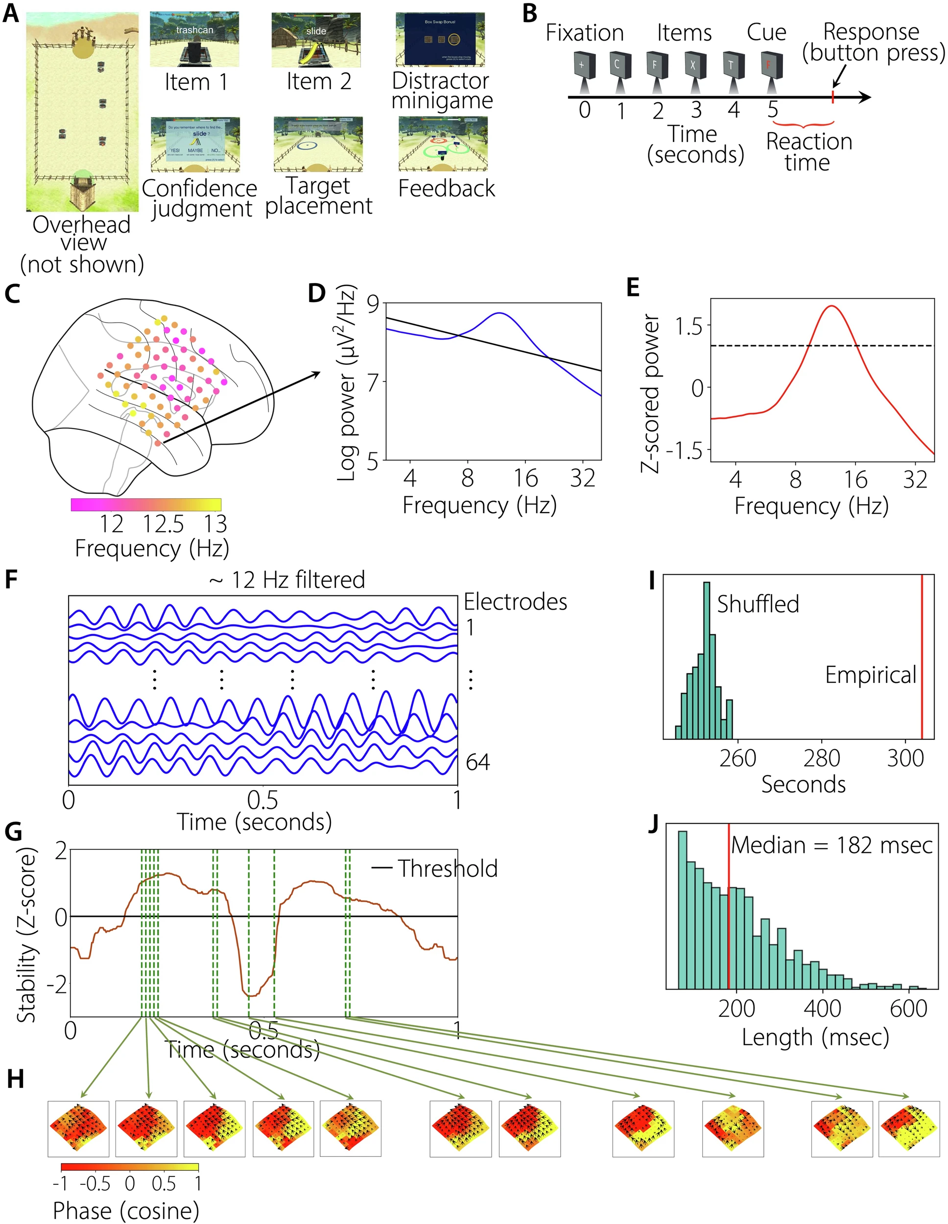
In the spatial task, participants navigated through a virtual environment containing treasure chests. Objects appeared at some of the chests, and participants later had to recall where those objects had been located. The researchers analyzed several stages of each trial, including encoding, navigation, confidence judgments, retrieval, distraction and feedback.
The verbal task used the classic Sternberg working-memory paradigm. Participants viewed sequences of one to three English letters and silently held them in memory before deciding whether a subsequent probe letter had appeared in the list. Across the task, participants viewed an 8- or 16-letter subset, and their mean accuracy was about 92 percent.
The spatial-memory task was less accurate, with mean location-recall accuracy of about 41 percent. The researchers analyzed short segments of neural activity from the different task periods rather than treating an entire trial as a single neural state.
The waves lasted only briefly
The analysis first identified narrowband oscillations in each electrode. The researchers examined frequencies from 3 to 40 hertz and looked for peaks above the background 1/f pattern in the power spectrum.
Oscillations were especially common in the theta, alpha and beta ranges. About 86 percent of all electrodes showed a narrowband oscillation in at least one of those ranges. Some electrode grids contained peaks in both the lower-frequency theta/alpha range and the higher-frequency beta range, suggesting that multiple oscillatory patterns could coexist in the same cortical region.
The researchers then estimated the instantaneous phase of each oscillation and examined how that phase varied across neighboring electrodes. A localized circular-linear regression provided an estimate of the direction and strength of propagation at each moment.
Rather than assuming that a wave remained stable throughout a trial, they looked for short periods during which its spatial pattern remained relatively consistent. These stable wave epochs lasted about 80 to 180 milliseconds on average.
The stability of those epochs was tested against shuffled data. The resulting tests were statistically significant, with all reported values below 0.001. Stability periods were shorter for higher-frequency oscillations than for lower-frequency oscillations: the median was 107 milliseconds versus 134 milliseconds, respectively, with p = 0.002 in a Mann-Whitney U test.
The waves also had measurable propagation speeds. Across the recordings, speeds ranged from about 0.3 to 3.3 meters per second. Higher-frequency waves traveled faster, with a median speed of 1.1 meters per second for 12–26 Hz oscillations compared with 0.5 meters per second for 5–12 Hz oscillations, a difference with p < 0.001.
For the main analysis, the researchers retained stable epochs longer than 25 sampling points. That corresponded to at least 50 milliseconds in the spatial-memory recordings and 62.5 milliseconds in the verbal-memory recordings. About 48 percent of the detected stable epochs met this criterion, producing roughly four usable stable epochs per trial.
A map of many different wave shapes
The researchers next used complex independent component analysis, or CICA, to separate the different spatial patterns contained in the traveling waves.
For each stable epoch, they represented the direction and strength of propagation at each electrode as a two-dimensional quantity. CICA then separated the combined activity into independent spatial patterns, which the researchers called modes. The weight assigned to a mode at a particular epoch indicated how strongly that pattern contributed at that time.
The resulting modes revealed much more than simple plane waves.
The researchers classified the patterns into four broad categories: planar, rotational, concentric and complex. Rotational waves included clockwise and counterclockwise patterns. Concentric waves could either expand outward from a source or contract inward toward a sink. Complex waves had statistically robust spatial propagation but did not meet the criteria for the other three categories. Some of those complex patterns contained smaller regions with planar, rotational or concentric behavior.
The analysis found multiple independent spatial patterns within individual electrode clusters. On average, each cluster had six significant modes. The first three modes explained roughly 34 percent, 19 percent and 10 percent of the variance, respectively, while the first six together explained more than 80 percent.
The researchers also tested whether CICA could recover known wave patterns in simulations. They introduced simulated planar, rotational, source and complex waves, added noise, and tested the algorithm at different signal-to-noise ratios. They also simulated combinations of multiple wave types.
At higher signal-to-noise ratios, the algorithm produced near-perfect reconstructions of the simulated patterns. Additional simulations using spatially shifted patterns tested whether CICA would incorrectly identify a particular wave shape simply because the data had similar spatial autocorrelation. In those tests, the shifted patterns did not reproduce the original wave structure.
A further simulation mixed trials containing waves with trials containing no wave. The algorithm correctly distinguished the two, reaching near-perfect detection at signal-to-noise ratios above about 15 decibels for rotational waves and above about 20 decibels for a combination of planar, source and rotational waves.
Straight waves remained common, but they were not alone
Planar waves were the most prominent category overall, particularly at lower frequencies.
In the spatial-memory and verbal-memory tasks, planar waves had a median strength of about 59 percent across task periods at lower frequencies. At higher frequencies, planar waves remained the most prevalent overall, with a median strength of about 43 percent, but rotational waves became more common, reaching a median strength of about 37 percent across tasks. The increase in rotational waves at higher frequencies was especially evident in the spatial-memory task.
The distribution also differed between the two hemispheres. Planar waves were stronger in the right hemisphere, where they accounted for about 64 percent of wave strength, compared with about 42 percent in the left hemisphere.
The balance of other wave types also differed between tasks. In the left hemisphere, rotational patterns were more common in the spatial-memory task, whereas concentric patterns were more common in the verbal-memory task. The right hemisphere showed statistically similar patterns between the two tasks.
The researchers did not find evidence that rotational waves preferred one of two possible large-scale directions. In the spatial-memory task, they identified seven temporal-to-parietal-to-frontal waves and nine in the opposite direction, a difference that was not statistically significant. In the verbal task, there were 12 and 15, respectively, again without a significant preference.
The concentric waves did show a difference between the tasks. In the spatial-memory recordings, 78 percent of the identified concentric waves were outward-moving sources, with seven sources and two sinks. In the verbal-memory recordings, 68 percent were inward-moving sinks, with 15 sinks and seven sources. The difference in the types of concentric waves between the tasks was statistically significant at p < 0.02.
These observations describe the spatial patterns present in the recordings. The researchers proposed possible functional explanations for them, but those explanations were not directly demonstrated by the recordings.
Wave patterns changed with different stages of memory
The clearest behavioral difference emerged when the researchers compared the wave patterns between different stages of the spatial-memory task.
In individual electrode clusters, the same underlying modes could change their direction, their strength, or both as participants moved from one stage of a trial to another.
In one example, a planar wave was nearly absent during navigation and distraction but was strongly represented during other stages. Its propagation direction also differed during retrieval compared with encoding, confidence and feedback. Another mode changed direction between memory stages and was strongest during retrieval and feedback.
This pattern was not restricted to the example. All 13 oscillation clusters analyzed in the spatial-memory task showed statistically significant shifts in traveling-wave direction or strength between behavioral stages.
The researchers used MANOVA to compare the complex CICA weights associated with different behavioral states. Because the weights contain both real and imaginary components, the statistical model considered both. Multiple comparisons were controlled using false-discovery-rate correction.
They also repeated the analysis after shuffling behavioral labels. Those shuffled analyses continued to distinguish the empirical results from the surrogate distributions, supporting the conclusion that the observed relationships between wave patterns and behavioral states were not simply produced by the analysis procedure.
The patterns could distinguish individual memory items
The researchers then asked whether the traveling waves reflected not only broad stages such as encoding or retrieval but also the particular item being held in memory.
In the verbal task, different English letters were associated with different propagation patterns in many of the electrode clusters. One example showed a planar wave traveling posteriorly for most of the letters but behaving differently for the letter G. Another mode propagated in one direction for G and Q and in the opposite direction for D and J.
Across the dataset, 13 of 26 oscillation clusters showed traveling waves that changed direction or strength depending on the individual letter being presented.
The statistical analysis therefore linked differences in wave patterns with both broad behavioral stages and more specific memory representations.
Spirals, sources and sinks also carried behavioral information
The more complex wave types were not merely occasional visual features.
Rotational waves were stable at the individual-epoch level and could distinguish behavioral states. They appeared across frontal, temporal and parietal regions in both memory tasks. In the spatial-memory task, rotational-wave strength was higher during the distractor period than during the other task periods, particularly in the left hemisphere, with χ²(5) = 67.0 and p < 0.001.
Concentric and complex waves likewise distinguished behavioral states across regions and tasks.
Complex-wave strength was significantly higher during the distractor phase than during the other task periods across frequencies and hemispheres, with χ²(5) = 43.2 and p < 0.001. The same pattern was not observed for concentric waves, for which χ²(5) was below 19.6 and p was greater than 0.01.
At the same time, the proportion of statistically significant modes did not differ significantly among the four wave categories or among the different modes. The researchers therefore found evidence that the different wave shapes were behaviorally informative without finding that one category simply contained a greater proportion of statistically significant modes.
The wave patterns could be decoded from the recordings
To test the behavioral relationship another way, the researchers trained multilayer neural networks to distinguish pairs of behavioral states from the CICA-derived wave features.
The networks were trained separately for each oscillation cluster, using five-fold cross-validation. Each model was trained on 80 percent of the data and tested on the remaining 20 percent, with the data balanced between the behavioral states being compared.
In the spatial-memory task, the neural networks could decode behavioral states at both individual-cluster and group levels. Group-level decoding was significantly above chance, with p < 0.001 in a one-sided sign test. The researchers found that some pairs of states were easier to distinguish than others.
Navigation and distraction were generally distinguishable from the other states but were relatively difficult to distinguish from each other. The researchers interpreted this as evidence that these two states had more similar traveling-wave patterns than the other behavioral states.
The same approach could decode the identity of individual letters in the verbal-memory task at the group level, with p < 0.001. Among the letters tested, H was often the most readily decoded, followed by J and Q.
Decoding accuracy also tracked how different the corresponding wave patterns were. Pairs of behavioral states with more dissimilar CICA patterns tended to be easier for the neural network to distinguish. The correlations between decoding accuracy and wave-pattern dissimilarity were statistically significant across the clusters, with all reported p values below 0.001 in the spatial-memory analysis.
Importantly, the researchers tested whether differences in oscillation frequency alone could account for the behavioral effects. Peak frequencies did not differ significantly among the task periods in the spatial-memory task or among the letters in the verbal-memory task, with all p values greater than 0.05. Neural networks also could not decode the behavioral states from peak frequency alone.
A similar control using the correlation between filtered and raw signals found that signal-to-noise differences could not account for the decoding results. The median correlation between filtered and raw recordings was about 0.6 across clusters, with all p values below 0.001, and decoding based on those correlations did not distinguish behavioral states.
What the recordings do not establish
The recordings show that traveling-wave patterns change with memory-related behavioral states, but they do not by themselves establish what generates those patterns or exactly what function each wave type performs.
The researchers discuss several possible mechanisms. Models of locally coupled neural oscillators suggest that waves can emerge from interactions among nearby neurons, conduction delays and differences in local oscillation frequencies. Within such models, spatial frequency gradients could generate planar, source or sink waves, while particular spatial arrangements could produce spirals.
Those mechanisms are presented as possible explanations rather than demonstrated causes of the observed human recordings.
The same qualification applies to the authors’ interpretation of source waves in spatial memory and sink waves in verbal memory. They propose that outward-moving sources might be associated with information spreading from local assemblies and that inward-moving sinks might reflect convergence onto particular regions. The recordings establish the differing prevalence of these patterns between the tasks, but they do not directly demonstrate that sources broadcast memory information or that sinks maintain verbal items.
The researchers also note that the waves sometimes appeared to travel through sulci and gyri and even across the Sylvian fissure. They discuss stable electric fields and possible subcortical, including thalamic, generation as potential explanations. Direct evidence from denser three-dimensional recordings would be needed to determine how such waves are generated and propagated through the full three-dimensional structure of the brain.
There is also a limitation in identifying rotational waves from the electrode arrays used here. Some of the grids were relatively small, including 6-by-4 and 8-by-4 arrangements. A planar pattern detected across such a limited area could represent only part of a larger rotational wave. The researchers therefore note that their method could have undercounted rotational waves. They suggest that larger ECoG arrays across more individuals will be needed to assess the prevalence and functional role of these patterns more accurately.
Another important source of uncertainty is the substantial variation in wave direction and strength among electrode clusters, brain regions and individuals. The researchers suggest that anatomical differences, as well as differences in local oscillation frequency and amplitude, could contribute to this variability. These possibilities remain to be tested.
The study therefore establishes a detailed spatial description of traveling waves during two forms of human memory processing. It shows that the waves are not limited to a single direction of propagation: their recorded patterns include planar, rotational, concentric and complex forms, and those patterns vary with both the stage of a memory task and, in the verbal task, the identity of individual remembered items. The neural-network analyses further showed that these wave patterns contained enough information to distinguish behavioral states in the recorded participants.
The study was published in Nature Communications.