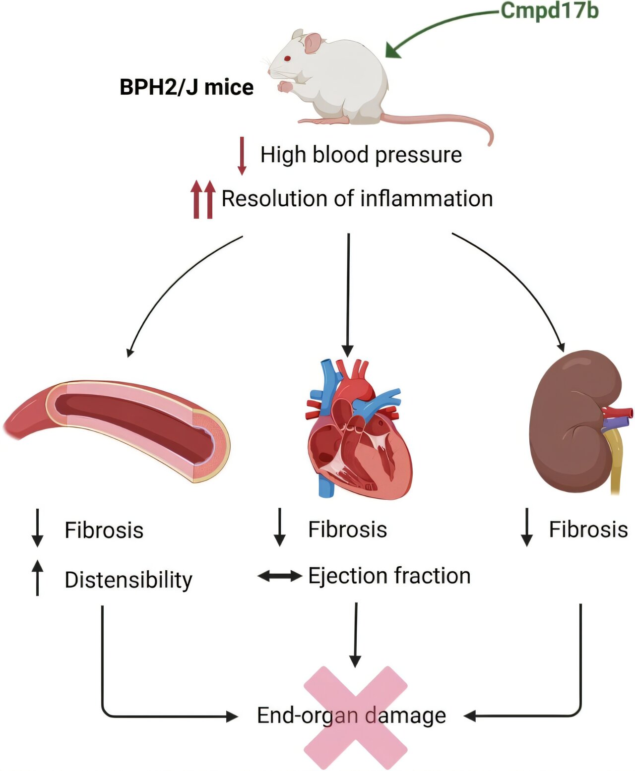
Cmpd17b lowered blood pressure and reduced fibrosis in genetically hypertensive mice, with the clearest effects seen in the kidneys. The four-week treatment also reduced collagen buildup in the aorta and heart, while changing the activity of genes linked to inflammation and tissue remodeling.
The researchers tested Cmpd17b in BPH/2J mice, a genetic mouse model of hypertension characterized by increased sympathetic nervous system activity. The animals develop hypertension early in life and show changes involving the kidneys, blood vessels and heart.
The study included 12-week-old male mice from two strains. BPH/2J mice had established high blood pressure, while BPN/3J mice served as normotensive controls. The animals received either Cmpd17b or a vehicle treatment every day for four weeks.
Cmpd17b is a synthetic ligand that activates formylpeptide receptors, or FPRs. These receptors are involved in regulating and resolving inflammation and are found on several types of cells, including immune cells and cells in the blood vessels, kidneys and heart.
The researchers had previously found that Cmpd17b could reduce cardiovascular damage in other experimental models of hypertension. This study asked whether the compound could also act after hypertension was already established in BPH/2J mice.
Blood pressure was measured continuously with implanted radiotelemetry devices. The researchers also examined the heart, blood vessels and kidneys using imaging, tissue analysis and measurements of gene activity.
Blood pressure fell in the hypertensive mice
At the start of the experiment, mean arterial pressure was 19% higher in BPH/2J mice than in BPN/3J mice over a 24-hour period.
After four weeks of Cmpd17b treatment, mean arterial pressure in the hypertensive BPH/2J mice had fallen by 6 mmHg from baseline. No change from baseline was observed in normotensive BPN/3J mice treated with Cmpd17b.
The researchers also reported reductions in systolic and diastolic arterial pressure in Cmpd17b-treated BPH/2J mice.
The blood-pressure effect was not simply an immediate response to each injection. Measurements taken 30 to 60 minutes after injections showed no short-term blood-pressure-lowering effect from Cmpd17b. The longer-term reduction therefore occurred without an obvious acute vasodilator response.
The effect was also stronger during the mice’s dark, active period, when blood pressure is highest in this model.
The treatment improved several measures of vascular remodeling
The hypertensive mice had less distensible aortas and thicker arterial walls than the normotensive mice.
After treatment with Cmpd17b, aortic distensibility was 37% greater and aortic wall thickness was 22% lower in BPH/2J mice than in vehicle-treated hypertensive mice.
The amount of collagen in the aorta was also lower after Cmpd17b treatment. Vehicle-treated BPH/2J mice had about twice as much aortic collagen deposition as BPN/3J mice, while Cmpd17b reduced collagen deposition in the hypertensive mice.
Some changes in the mesenteric artery were also observed. The hypertensive mice had thicker mesenteric artery walls, and this measure tended to be lower after Cmpd17b treatment.
However, not every vascular abnormality improved. The hypertensive mice had impaired acetylcholine-induced relaxation in mesenteric arteries, and Cmpd17b did not improve that response. Other measurements of the structure and passive mechanical properties of the mesenteric and femoral arteries did not differ among the groups.
These results meant that the vascular effects were selective rather than a general correction of every measured vascular abnormality.
Kidney fibrosis showed the strongest response
The clearest structural effect appeared in the kidneys.
Vehicle-treated BPH/2J mice had more interstitial collagen deposition in the kidney than normotensive BPN/3J mice. Cmpd17b reduced this interstitial collagen accumulation in the hypertensive mice.
The treatment did not change kidney weight in either mouse strain. Kidney weight relative to tibia length was lower in BPH/2J mice than in BPN/3J mice, regardless of treatment.
The molecular results also pointed toward changes in the kidney.
RNA sequencing found 1,712 genes that were significantly different between untreated hypertensive and normotensive mice in the kidney. In the left ventricle, 1,336 genes were significantly different.
Most of these changes were specific to one tissue. There were 1,260 genes uniquely altered in the kidney and 884 uniquely altered in the left ventricle. Another 452 genes were significantly changed in both tissues.
When the researchers compared Cmpd17b-treated and vehicle-treated hypertensive mice, they found no differentially expressed genes in the left ventricle under their RNA-sequencing criteria. In the kidney, however, 34 genes were upregulated and five were downregulated after Cmpd17b treatment.
Those 39 genes were not significantly affected by Cmpd17b in the normotensive mice.
Two genes received particular attention. Serpina1e was strongly increased in the kidneys of hypertensive mice and decreased after Cmpd17b treatment. Igfbp2 also showed a reversal in expression, although the change after treatment did not meet the study’s statistical threshold for significance.
Pathway analysis identified reduced activity in several inflammatory and immune-related pathways in the kidneys of Cmpd17b-treated hypertensive mice. These included chemokine signaling, tumor necrosis factor signaling, and complement and coagulation cascades.
Gene measurements supported the kidney findings
The researchers used RT-qPCR to examine selected genes identified through the tissue analyses.
In the kidneys of vehicle-treated BPH/2J mice, expression of S100a9, an inflammatory gene, and Ctgf, a gene associated with fibrosis, was higher than in BPN/3J mice. Both were reduced in the kidneys of Cmpd17b-treated hypertensive mice.
The researchers also examined the left ventricle. Although RNA sequencing did not identify significant treatment-related gene changes there, RT-qPCR showed that S100a9 and Mmp2 were elevated in vehicle-treated BPH/2J mice and lower after Cmpd17b treatment.
The results therefore provided molecular evidence of changes related to inflammation and tissue remodeling in both organs, although the kidney showed a much broader transcriptional response.
Heart fibrosis decreased without improved heart function
The BPH/2J mice did not show overt cardiac hypertrophy in this experiment. Heart and left-ventricle weight were comparable among the groups, and cardiomyocyte area was also similar.
The hypertensive mice did have more collagen in the left ventricle. Levels of collagen I, collagen III and total collagen were higher than in normotensive mice.
Cmpd17b reduced all three measures of left-ventricular collagen deposition in hypertensive mice.
But the structural improvement did not translate into a detectable improvement in cardiac function. Echocardiography showed reduced fractional shortening in BPH/2J mice compared with BPN/3J mice, while ejection fraction was not significantly different between individual groups. Cmpd17b did not improve cardiac function.
The treatment therefore produced evidence of reduced cardiac fibrosis without correcting the measured functional changes.
The blood-pressure effect was not explained by sympathetic activity
Because BPH/2J hypertension is strongly associated with sympathetic nervous system overactivity, the researchers examined whether Cmpd17b changed an indirect measure of sympathetic activity called mid-frequency mean arterial pressure power.
As expected, this measure was higher in BPH/2J mice at baseline.
But both Cmpd17b and the vehicle treatment reduced the measure in hypertensive mice. Because the vehicle produced a similar reduction, the researchers concluded that the change could not be attributed specifically to Cmpd17b.
The reduction in blood pressure produced by Cmpd17b was greater than that produced by the vehicle. The researchers therefore concluded that the blood-pressure effect was not explained by the modest reduction in sympathetic activity associated with the vehicle.
Cmpd17b also did not change renal Ren1 expression, another measure associated with sympathetic activity in this mouse model.
These findings differed from earlier work in an angiotensin II-induced model of hypertension, in which Cmpd17b had reduced sympathetic outflow. The researchers therefore concluded that the mechanism operating in BPH/2J mice was different.
The treatment also acted without changing the renin-angiotensin system
The researchers examined whether Cmpd17b changed the renin-angiotensin-aldosterone system, which contributes to blood-pressure regulation.
Ren1 expression was higher in vehicle-treated BPH/2J mice than in BPN/3J mice, consistent with previous observations. Cmpd17b did not change Ren1 or Agt expression.
The compound also did not change the blood-pressure response to angiotensin II infusion.
The researchers therefore considered it unlikely that Cmpd17b lowered blood pressure by changing the renin-angiotensin system or sensitivity to angiotensin II.
The researchers linked the tissue effects to inflammation and remodeling
Taken together, the results showed a pattern across the major organs examined.
In hypertensive mice, Cmpd17b lowered blood pressure, improved aortic distensibility, reduced aortic wall thickness and decreased collagen deposition. In the kidney, it reduced interstitial fibrosis and changed a set of genes associated with inflammation and tissue remodeling. In the heart, it reduced collagen deposition and Mmp2 expression but did not improve measured cardiac function.
The researchers concluded that the effects of Cmpd17b in established neurogenic hypertension were primarily associated with anti-inflammatory and tissue-remodeling mechanisms rather than direct changes in sympathetic activity.
The strongest effects were observed in the kidney and vasculature.
The study also found that these tissue changes occurred alongside only a moderate reduction in blood pressure. The researchers noted that their experiments could not completely separate effects caused by lower blood pressure from effects directly produced by Cmpd17b in the tissues.
The study has important limits
The experiment used only male mice. The researchers said this was done to reduce biological variability in the initial proof-of-concept study, but they noted that future work should include female mice to examine possible sex-specific differences.
The study also focused on structural and molecular measurements rather than direct measures of kidney function. Glomerular filtration rate was not measured.
Another limitation was that endothelial-mediated vascular relaxation remained impaired despite treatment. The researchers noted that the existing vascular damage may have exceeded the capacity of Cmpd17b to repair it and suggested that future studies should examine earlier treatment or combinations with vascular repair agents.
The researchers also cautioned against making broad conclusions about FPR activation itself. FPR signaling depends on the ligand, and different FPR activation patterns can produce different effects. The study specifically tested Cmpd17b rather than establishing that all FPR activation would have the same effects in hypertension.
The experiments were conducted in mice, so the findings do not establish that Cmpd17b is effective for treating hypertension in people.
The study was published in Communications Biology.