From the moment you wake and feel the softness of your pillow to the instant you drift into dreams, your brain orchestrates every sensation, thought, and emotion. This three-pound organ, nestled within the protective embrace of your skull, is the seat of consciousness, the engine of creativity, and the archive of your memories. It interprets the world, plans your actions, and gives rise to the rich tapestry of experiences that define who you are. Yet despite centuries of inquiry, the brain remains the most complex object in the known universe. In this article, we embark on an epic voyage into its mysterious depths, exploring how billions of cells work in concert to transform electrical impulses into thoughts, feelings, and actions.
Our journey will take us from the microscopic architecture of neurons and synapses to the breathtaking panorama of brain networks. We will delve into developmental processes that shape our minds, the astonishing plasticity that endows us with learning abilities, and the neural underpinnings of consciousness itself. Along the way, we will glimpse how modern technologies—such as functional imaging and computational modeling—are illuminating the brain’s labyrinthine circuits, and how the study of brain disorders is teaching us about normal brain function. Ultimately, we aim not only to understand how the brain works but also to appreciate its elegance, resilience, and the profound mystery that still surrounds it.
Anatomy of the Brain: The Landscape Within
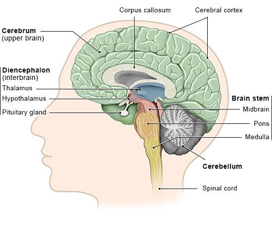
The human brain can be conceptually divided into several major regions, each with distinct structures and functions, yet all seamlessly integrated. On the outer surface lies the cerebral cortex, a wrinkled sheet of gray matter rippling across the brain’s hemispheres. These convolutions—gyri (ridges) and sulci (grooves)—increase surface area, housing roughly twenty billion neurons. Beneath the cortex resides the white matter, composed of bundled axons that interconnect distant brain regions. At the core, deeper structures such as the thalamus, hypothalamus, basal ganglia, and limbic system regulate fundamental processes like sensation relay, hormonal balance, movement, and emotion.
The hindbrain, including the brainstem and cerebellum, lies beneath the cerebrum and connects directly to the spinal cord. The brainstem—formed by the midbrain, pons, and medulla—governs vital autonomic functions: breathing, heart rate, blood pressure, and sleep–wake cycles. The cerebellum, with its own dense network of neurons, fine-tunes movement, balance, and certain cognitive tasks like language and attention.
Despite these regional distinctions, the brain operates as a harmonious whole. Information flows continuously: sensory data from your eyes and ears stream through relay stations, ultimately reaching the cortex for interpretation. Motor commands from the cortex descend through the brainstem to the spinal cord, orchestrating muscle contractions. Meanwhile, the limbic system modulates emotional context, and neuromodulators like dopamine and serotonin adjust the brain’s overall state. This intricate choreography unfolds in real time, enabling you to perceive, decide, and act in a constantly changing world.
Neurons: The Fundamental Units
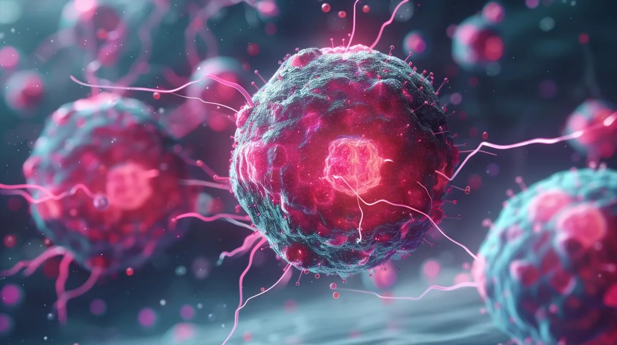
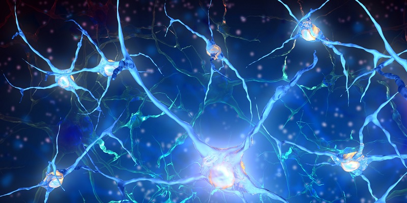
At the heart of brain function lie neurons—specialized cells exquisitely adapted for rapid communication. A typical neuron features a cell body (soma), numerous branching dendrites that receive incoming signals, and a long axon that transmits signals to other cells. At the axon terminal, the neuron releases neurotransmitters—chemical messengers that bridge the gap (synapse) to the next neuron’s dendrite or cell body.
Neurons come in diverse morphologies and types, each tailored to specific roles. Excitatory neurons, often using glutamate as their neurotransmitter, promote the firing of their targets. Inhibitory neurons, frequently releasing GABA, temper overactivity and sculpt precise patterns of brain activity. Other specialized neurons release modulators like dopamine, norepinephrine, and acetylcholine, influencing mood, arousal, and attention.
A neuron generates electrical signals through the movement of ions across its membrane. When excitatory inputs push the membrane potential past a threshold—typically around –55 millivolts—the neuron fires an action potential. This rapid depolarization travels along the axon at speeds ranging from a few meters per second up to 120 meters per second in myelinated fibers. Upon reaching the synapse, the action potential triggers the influx of calcium ions, leading to neurotransmitter release. The transmitted signal then influences the receiving neuron, continuing the chain of communication.
Synapses and Circuits: Forging Connections
The brain’s power emerges not from individual neurons but from the colossal network of synaptic connections among them. Each neuron may form thousands of synapses, creating trillions of points of contact. These synapses can be excitatory or inhibitory, and their strengths—governed by receptor density, neurotransmitter release probability, and other factors—determine the flow of information through neural circuits.
Synaptic plasticity—the dynamic adjustment of synaptic strength—is the cellular foundation of learning and memory. When two neurons fire together repeatedly, their connection strengthens, a phenomenon known as long-term potentiation (LTP). Conversely, long-term depression (LTD) weakens connections when activity patterns dictate. These processes modify the topology and weightings of neural networks, enabling the brain to encode new experiences, skills, and associations.
At a larger scale, groups of interconnected neurons form functional circuits. In the visual system, for example, signals travel from the retina to the lateral geniculate nucleus of the thalamus, then to primary visual cortex, and on to higher-order regions that process color, motion, and form. Motor circuits integrate information from the cortex, cerebellum, and basal ganglia to plan and execute movements. Emotional circuits within the limbic system evaluate stimuli, generate feelings, and guide decision-making.
Understanding these circuits has been greatly advanced by modern techniques such as optogenetics—where light-sensitive proteins control neuronal firing—and connectomics, which maps the brain’s wiring diagram at ever finer resolution. While we still lack a complete wiring map of the human brain, these tools are revealing the principles by which circuits generate perception, thought, and behavior.
Development and Wiring: From Embryo to Adult
The formation of the human brain begins early in embryonic development, when a sheet of neural plate folds to form the neural tube. Neural progenitor cells within this tube proliferate rapidly, giving rise to billions of neurons and glial cells. Guided by molecular cues, newborn neurons migrate to their destined locations in the cortex, cerebellum, and other regions.
Neuronal migration is followed by axon guidance, where growth cones at axon tips follow chemical gradients to reach target cells. Along the way, axons form provisional synapses. Early in life, the brain generates more synapses than it ultimately needs. This exuberant connectivity is pruned by experience—unused synapses retract, while those reinforced by activity persist. This process of synaptic pruning is most intense during critical periods, such as early childhood for vision and language, shaping the brain’s capabilities.
Glial cells—astrocytes, oligodendrocytes, and microglia—play crucial roles in development. Astrocytes provide metabolic support, regulate extracellular ion concentrations, and participate in synapse formation. Oligodendrocytes wrap axons in myelin, accelerating electrical conduction. Microglia, the brain’s immune cells, sculpt synapses and respond to injury or infection.
By adolescence, the brain undergoes a second wave of remodeling. The prefrontal cortex—responsible for judgment, impulse control, and planning—continues to mature into the mid-twenties. This protracted development underlies the shifts in risk-taking, emotional regulation, and social cognition characteristic of teenage years and early adulthood.
Plasticity: The Brain’s Remarkable Adaptability
One of the most astounding features of the human brain is its plasticity—its lifelong capacity to adapt structurally and functionally. While early critical periods set the foundation, the brain remains malleable throughout life, enabling learning, recovery from injury, and adaptation to new environments.
Experience-dependent plasticity manifests in many forms. Skill acquisition—learning to play an instrument or speak a new language—strengthens relevant neural circuits. London taxi drivers, who memorize the city’s labyrinthine streets, exhibit increased volume in the hippocampus, a region crucial for spatial memory. In individuals who lose a limb, brain areas once devoted to that limb can be repurposed to process input from adjacent regions, illustrating the brain’s capacity for functional reorganization.
At the cellular level, plasticity involves not only synaptic changes but also structural modifications: the growth of new dendritic spines, the generation of new neurons in certain regions (notably the hippocampus), and alterations in myelination. Even in older adults, engaging in challenging cognitive tasks can stimulate neural growth and maintain cognitive health.
Plasticity also underlies recovery from brain injuries. After a stroke, surviving neurons can form new connections to compensate for lost function. Rehabilitation therapies—physical, occupational, or speech therapy—leverage this plasticity by repeatedly engaging affected circuits, promoting the reestablishment of skills.
The Neural Basis of Perception and Action
At every moment, your brain constructs a model of the external world based on sensory input. Photons entering your eyes trigger cascades of activity across retinal photoreceptors, bipolar cells, and ganglion cells; action potentials then travel via the optic nerve to the visual cortex. There, hierarchical processing extracts edges, colors, motion, and ultimately object identity and spatial relationships.
Auditory information travels from hair cells in the cochlea through brainstem nuclei to the auditory cortex, enabling you to perceive pitch, timbre, and location. Smell and taste rely on chemoreceptors that detect molecular signatures, evoking rich perceptions and emotional memories. The somatosensory system transforms tactile, temperature, and pain signals into conscious awareness of your body and its interactions with the environment.
When you decide to move, neural commands originate in motor areas of the cortex, are refined through basal ganglia and cerebellar loops, and descend via spinal interneurons to motor neurons. These, in turn, trigger muscle fibers to contract, executing precise gestures. Sensory feedback continuously informs the brain about the outcome, allowing moment-to-moment correction and fluidity.
This sensorimotor integration exemplifies the brain’s predictive nature. Rather than passively registering stimuli, the brain actively anticipates sensory consequences of actions. Discrepancies between expected and actual feedback drive learning and refine motor control—a principle at the heart of theories like predictive coding.
Consciousness and Cognition: The Pinnacle of Brain Function
Perhaps the greatest mystery of all is consciousness—the subjective awareness of self and world. Despite decades of research, no single brain region or mechanism has been singled out as a “consciousness center.” Instead, consciousness appears to arise from the dynamic interplay of widespread networks, including fronto-parietal systems, thalamocortical loops, and default mode and salience networks.
Higher cognitive functions—attention, working memory, decision-making, language, and social cognition—depend on the coordinated activity of distributed circuits. The prefrontal cortex, with its extensive connections, plays a crucial role in executive functions: planning, inhibiting inappropriate responses, and flexibly switching between tasks. The language network, anchored in Broca’s and Wernicke’s areas, enables us to comprehend and produce speech. The theory of mind—our ability to attribute mental states to others—emerges from interactions among medial prefrontal cortex, temporal-parietal junction, and other regions.
Recent advances in neuroimaging and electrophysiology have begun to reveal the temporal and spatial dynamics of these processes. Techniques like magnetoencephalography (MEG) and intracranial recordings capture the millisecond-by-millisecond choreography of neuronal ensembles. Functional MRI (fMRI) maps brain regions that coactivate during specific tasks, offering insights into cognitive architectures. Yet, translating these observations into a cohesive theory of consciousness remains an ongoing quest.
Disorders of the Brain: Windows into Normal Function
When the brain malfunctions, the resulting disorders can be devastating. Studying these conditions has shed light on normal brain operations. Parkinson’s disease, characterized by tremors and movement rigidity, stems from degeneration of dopamine-producing neurons in the substantia nigra. Its discovery highlighted the role of dopamine in motor control and reward processing.
Alzheimer’s disease, marked by memory loss and cognitive decline, involves accumulation of amyloid-beta plaques and tau tangles, leading to neuronal death. Research into Alzheimer’s has illuminated mechanisms of protein aggregation, synaptic loss, and neuroinflammation. Epilepsy, autism spectrum disorder, schizophrenia, depression, and many other conditions each offer unique insights into the neural substrates of cognition, emotion, and behavior.
Advances in genetics have identified risk genes and pathways implicated in these disorders. Gene-editing tools like CRISPR promise new avenues for understanding gene–brain relationships. Meanwhile, neurostimulation techniques—deep brain stimulation, transcranial magnetic stimulation, and focused ultrasound—are emerging as therapies that modulate dysfunctional circuits.
Mapping the Brain: Tools of Discovery
Our understanding of the brain has advanced hand in hand with technological innovation. Early neuroscientists relied on postmortem dissections and lesion studies, correlating brain injuries with behavioral deficits. The advent of the electron microscope revealed the ultrastructure of synapses and organelles. Electrophysiological recordings—single-cell and population-level—deciphered neuronal signaling in unprecedented detail.
In recent decades, imaging methods revolutionized neuroscience. MRI and CT scans provided noninvasive windows into brain anatomy. Functional imaging techniques—fMRI, PET, and SPECT—allowed visualization of metabolic and hemodynamic correlates of neural activity. Diffusion tensor imaging (DTI) maps white-matter tracts, unveiling the brain’s wiring.
Optical methods, like two-photon microscopy, enabled imaging of living tissue at subcellular resolution. Genetically encoded calcium and voltage indicators report neuronal activity in real time. Optogenetics combines genetic targeting of light-sensitive proteins with optical stimulation, granting millisecond control over specific cell populations.
On the computational front, powerful models simulate neuronal networks, bridging the gap between microscopic mechanisms and behavior. The Human Brain Project and the BRAIN Initiative aim to integrate data across scales, from genes to cognition, forging a new era of systems neuroscience.
The Brain and Artificial Intelligence: Lessons and Inspirations
The quest to understand the brain has inspired artificial intelligence (AI), while AI in turn offers tools for decoding neural data. Artificial neural networks mimic certain features of biological networks—layers of interconnected units that adjust connection weights through learning rules inspired by synaptic plasticity. Deep learning architectures have revolutionized image and speech recognition, reinforcement learning, and natural language processing.
Yet biological brains remain far more efficient, flexible, and robust. They operate at a fraction of the energy consumption of AI systems and can learn from far fewer examples. Incorporating biological principles—sparse coding, recurrent connectivity, neuromodulation—into AI holds promise for more powerful algorithms. Conversely, AI methods like machine learning aid in analyzing vast brain-mapping datasets, identifying patterns that elude human observers.
The interface between brain and machine is also growing tangible. Brain–computer interfaces decode neural signals to control prosthetic limbs or communicate with paralyzed patients. Advances in neural implants and neural lace technologies raise profound possibilities—and ethical questions—about enhancing cognition or merging minds with machines.
Ethical and Philosophical Reflections
Probing the brain touches on deep philosophical questions about identity, free will, and the nature of consciousness. If our thoughts arise from electrochemical processes, what becomes of notions like soul or self? Neuroscience increasingly demonstrates how genetic and environmental factors shape personality, challenging ideas of absolute autonomy. Meanwhile, advances in neurotechnology raise ethical dilemmas: who owns neural data? Should we enhance memory or intelligence? How do we protect mental privacy?
Public engagement and interdisciplinary dialogue—uniting neuroscientists, ethicists, legal scholars, and the wider community—are vital as we navigate these frontiers. The brain’s complexity demands humility and responsibility, ensuring that discoveries serve human well-being and dignity.
Conclusion: The Ongoing Odyssey
Our expedition into the human brain has revealed an extraordinary organ of staggering complexity, adaptability, and mystery. From individual ion channels to vast networks generating thoughts, the brain exemplifies nature’s ingenuity. While we have uncovered fundamental principles of neuronal signaling, synaptic plasticity, and circuit dynamics, the deepest secrets of consciousness and cognition remain hidden.
The next decades promise remarkable strides: more complete connectomes, real-time recordings of millions of neurons, breakthroughs in treating neurological and psychiatric disorders, and ever-closer integration of brains with machines. As we delve further into this final frontier, each discovery will reshape our understanding of ourselves and our place in the cosmos.
In the end, studying the brain is not merely a scientific endeavor—it is an intimate journey into what makes us human. It is the pursuit of understanding how a tangle of cells gives rise to poetry, art, love, and the quest for knowledge itself. The brain, the universe’s most complex known structure, invites us to explore not just its mechanisms but the very essence of life and consciousness. And so, the odyssey continues.